


1PG8 | pdb_00001pg8
1PG8 | pdb_00001pg8
Crystal Structure of L-methionine alpha-, gamma-lyase
- PDB DOI: https://doi.org/10.2210/pdb1PG8/pdb
- Classification: LYASE
- Organism(s): Pseudomonas putida
- Mutation(s): No
- Deposited: 2003-05-28 Released: 2004-06-08
Experimental Data Snapshot
- Method: X-RAY DIFFRACTION
- Resolution: 2.68 Å
- R-Value Free: 0.236 (Depositor)
- R-Value Work: 0.182 (Depositor), 0.182 (DCC)
- R-Value Observed: 0.182 (Depositor)
Starting Model: experimental
View more details

This is version 1.3 of the entry. See complete history.
Literature
To be published.

Macromolecules
Find similar proteins by:
| 3D Structure
Entity ID: 1 | |||||
|---|---|---|---|---|---|
| Molecule | Chains | Sequence Length | Organism | Details | Image |
| Methionine gamma-lyase | 398 | Pseudomonas putida | Mutation(s): 0 EC: 4.4.1.11 (PDB Primary Data), 4.4.1.2 (UniProt) | 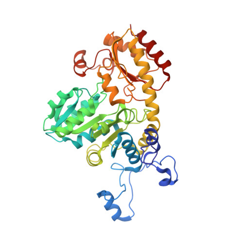 | |
UniProt | |||||
Entity Groups | |||||
| Sequence Clusters | 30% Identity50% Identity70% Identity90% Identity95% Identity100% Identity | ||||
| UniProt Group | P13254 | ||||
Sequence AnnotationsExpand | |||||
Reference Sequence | |||||
Small Molecules
| Ligands 3 Unique | |||||
|---|---|---|---|---|---|
| ID | Chains | Name / Formula / InChI Key | 2D Diagram | 3D Interactions | |
| PLP Download:Ideal Coordinates CCD File | F [auth A], O [auth B], S [auth C], W [auth D] | PYRIDOXAL-5'-PHOSPHATE C8 H10 N O6 P NGVDGCNFYWLIFO-UHFFFAOYSA-N |  | ||
| PEG Download:Ideal Coordinates CCD File | G [auth A] H [auth A] I [auth A] J [auth A] K [auth A] | DI(HYDROXYETHYL)ETHER C4 H10 O3 MTHSVFCYNBDYFN-UHFFFAOYSA-N |  | ||
| SO4 Download:Ideal Coordinates CCD File | E [auth A], N [auth B], R [auth C], V [auth D] | SULFATE ION O4 S QAOWNCQODCNURD-UHFFFAOYSA-L |  | ||
Experimental Data & Validation
Experimental Data
- Method: X-RAY DIFFRACTION
- Resolution: 2.68 Å
- R-Value Free: 0.236 (Depositor)
- R-Value Work: 0.182 (Depositor), 0.182 (DCC)
- R-Value Observed: 0.182 (Depositor)
Space Group: P 21 21 2
Unit Cell:
| Length ( Å ) | Angle ( ˚ ) |
|---|---|
| a = 152.83 | α = 90 |
| b = 154.64 | β = 90 |
| c = 80.79 | γ = 90 |
| Software Name | Purpose |
|---|---|
| SCALA | data scaling |
| AMoRE | phasing |
| REFMAC | refinement |
| CCP4 | data scaling |
Entry History
Deposition Data
- Released Date: 2004-06-08 Deposition Author(s): Allen, T.W., Sridhar, V., Prasad, G.S., Han, Q., Xu, M., Tan, Y., Hoffman, R.M., Ramaswamy, S.
Revision History (Full details and data files)
- Version 1.0: 2004-06-08
Type: Initial release - Version 1.1: 2008-04-29
Changes: Version format compliance - Version 1.2: 2011-07-13
Changes: Non-polymer description, Version format compliance - Version 1.3: 2023-08-16
Changes: Data collection, Database references, Derived calculations, Refinement description


