1PEV | pdb_00001pev

Crystal Structure of the Actin Interacting Protein from Caenorhabditis Elegans


Experimental Data Snapshot

  • Method: X-RAY DIFFRACTION
  • Resolution: 2.00 Å
  • R-Value Free: 
    0.234 (Depositor), 0.234 (DCC) 
  • R-Value Work: 
    0.197 (Depositor), 0.197 (DCC) 
  • R-Value Observed: 
    0.197 (Depositor) 

Starting Model: experimental
View more details

wwPDB Validation 3D Report Full Report

Validation slider image for 1PEV

This is version 1.4 of the entry. See complete history

Literature

Identification of functional residues on Caenorhabditis elegans actin-interacting protein 1 (UNC-78) for disassembly of actin depolymerizing factor/cofilin-bound actin filaments.

Mohri, K.Vorobiev, S.Fedorov, A.A.Almo, S.C.Ono, S.

(2004) J Biological Chem 279: 31697-31707

  • DOI: https://doi.org/10.1074/jbc.M403351200
  • Primary Citation Related Structures: 
    1NR0, 1PEV

  • PubMed Abstract: 

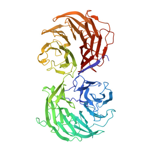
    Actin-interacting protein 1 (AIP1) is a WD40 repeat protein that enhances actin filament disassembly in the presence of actin-depolymerizing factor (ADF)/cofilin. AIP1 also caps the barbed end of ADF/cofilin-bound actin filament. However, the mechanism by which AIP1 interacts with ADF/cofilin and actin is not clearly understood. We determined the crystal structure of Caenorhabditis elegans AIP1 (UNC-78), which revealed 14 WD40 modules arranged in two seven-bladed beta-propeller domains. The structure allowed for the mapping of conserved surface residues, and mutagenesis studies identified five residues that affected the ADF/cofilin-dependent actin filament disassembly activity. Mutations of these residues, which reside in blades 3 and 4 in the N-terminal propeller domain, had significant effects on the disassembly activity but did not alter the barbed end capping activity. These data support a model in which this conserved surface of AIP1 plays a direct role in enhancing fragmentation/depolymerization of ADF/cofilin-bound actin filaments but not in barbed end capping.


  • Organizational Affiliation
    • Department of Pathology, Emory University, Atlanta, Georgia 30322, USA.

Macromolecule Content 

  • Total Structure Weight: 65.39 kDa 
  • Atom Count: 4,930 
  • Modeled Residue Count: 610 
  • Deposited Residue Count: 611 
  • Unique protein chains: 1

Macromolecules

Find similar proteins by:|  3D Structure
Entity ID: 1
MoleculeChains  Sequence LengthOrganismDetailsImage
Actin interacting protein 1611Caenorhabditis elegansMutation(s): 0 
Gene Names: UNC-78 OR C04F6.4
UniProt
Find proteins for Q11176 (Caenorhabditis elegans)
Explore Q11176 
Go to UniProtKB:  Q11176
Entity Groups
Sequence Clusters30% Identity50% Identity70% Identity90% Identity95% Identity100% Identity
UniProt GroupQ11176
Sequence Annotations
Expand
Reference Sequence

Experimental Data & Validation

Experimental Data

  • Method: X-RAY DIFFRACTION
  • Resolution: 2.00 Å
  • R-Value Free:  0.234 (Depositor), 0.234 (DCC) 
  • R-Value Work:  0.197 (Depositor), 0.197 (DCC) 
  • R-Value Observed: 0.197 (Depositor) 
Space Group: P 1 21 1
Unit Cell:
Length ( Å )Angle ( ˚ )
a = 40.66α = 90
b = 90.817β = 94.06
c = 76.983γ = 90
Software Package:
Software NamePurpose
CNSrefinement
HKL-2000data reduction
SCALEPACKdata scaling
CNSphasing

Structure Validation

View Full Validation Report


Entry History 

Revision History  (Full details and data files)

  • Version 1.0: 2003-07-01
    Type: Initial release
  • Version 1.1: 2008-04-29
    Changes: Version format compliance
  • Version 1.2: 2011-07-13
    Changes: Version format compliance
  • Version 1.3: 2021-02-03
    Changes: Structure summary
  • Version 1.4: 2023-08-16
    Changes: Data collection, Database references, Refinement description