1PC6 | pdb_00001pc6

Structural Genomics, NinB


Experimental Data Snapshot

  • Method: X-RAY DIFFRACTION
  • Resolution: 2.51 Å
  • R-Value Free: 
    0.294 (Depositor), 0.305 (DCC) 
  • R-Value Work: 
    0.234 (Depositor), 0.241 (DCC) 
  • R-Value Observed: 
    0.234 (Depositor) 

wwPDB Validation 3D Report Full Report

Validation slider image for 1PC6

This is version 1.4 of the entry. See complete history

Literature

Functional similarities between phage lambda Orf and Escherichia coli RecFOR in initiation of genetic exchange

Maxwell, K.L.Reed, P.Zhang, R.Beasley, S.Walmsley, A.R.Curtis, F.A.Joachimiak, A.Edwards, A.M.Sharples, G.J.

(2005) Proc Natl Acad Sci U S A 102: 11260-11265

  • DOI: https://doi.org/10.1073/pnas.0503399102
  • Primary Citation Related Structures: 
    1PC6

  • PubMed Abstract: 

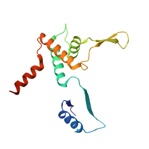
    Genetic recombination in bacteriophage lambda relies on DNA end processing by Exo to expose 3'-tailed strands for annealing and exchange by beta protein. Phage lambda encodes an additional recombinase, Orf, which participates in the early stages of recombination by supplying a function equivalent to the Escherichia coli RecFOR complex. These host enzymes assist loading of the RecA strand exchange protein onto ssDNA coated with ssDNA-binding protein. In this study, we purified the Orf protein, analyzed its biochemical properties, and determined its crystal structure at 2.5 angstroms. The homodimeric Orf protein is arranged as a toroid with a shallow U-shaped cleft, lined with basic residues, running perpendicular to the central cavity. Orf binds DNA, favoring single-stranded over duplex and with no obvious preference for gapped, 3'-tailed, or 5'-tailed substrates. An interaction between Orf and ssDNA-binding protein was indicated by far Western analysis. The functional similarities between Orf and RecFOR are discussed in relation to the early steps of recombinational exchange and the interplay between phage and bacterial recombinases.


  • Organizational Affiliation
    • Centre for Infectious Diseases, Wolfson Research Institute, University of Durham, Queen's Campus, Stockton-on-Tees TS17 6BH, United Kingdom.

Macromolecule Content 

  • Total Structure Weight: 33.5 kDa 
  • Atom Count: 2,170 
  • Modeled Residue Count: 274 
  • Deposited Residue Count: 292 
  • Unique protein chains: 1

Macromolecules

Find similar proteins by:|  3D Structure
Entity ID: 1
MoleculeChains  Sequence LengthOrganismDetailsImage
Protein ninB
A, B
146Lambdavirus lambdaMutation(s): 0 
UniProt
Find proteins for P03765 (Escherichia phage lambda)
Explore P03765 
Go to UniProtKB:  P03765
Entity Groups
Sequence Clusters30% Identity50% Identity70% Identity90% Identity95% Identity100% Identity
UniProt GroupP03765
Sequence Annotations
Expand
Reference Sequence

Experimental Data & Validation

Experimental Data

  • Method: X-RAY DIFFRACTION
  • Resolution: 2.51 Å
  • R-Value Free:  0.294 (Depositor), 0.305 (DCC) 
  • R-Value Work:  0.234 (Depositor), 0.241 (DCC) 
  • R-Value Observed: 0.234 (Depositor) 
Space Group: P 32 2 1
Unit Cell:
Length ( Å )Angle ( ˚ )
a = 76.776α = 90
b = 76.776β = 90
c = 107.39γ = 120
Software Package:
Software NamePurpose
CNSrefinement
d*TREKdata reduction
HKL-2000data scaling
CNSphasing

Structure Validation

View Full Validation Report


Entry History 

Deposition Data

Revision History  (Full details and data files)

  • Version 1.0: 2004-01-20
    Type: Initial release
  • Version 1.1: 2008-04-29
    Changes: Version format compliance
  • Version 1.2: 2011-07-13
    Changes: Version format compliance
  • Version 1.3: 2017-10-11
    Changes: Refinement description
  • Version 1.4: 2025-03-26
    Changes: Data collection, Database references, Derived calculations, Structure summary