1PBR | pdb_00001pbr

STRUCTURE OF 16S RIBOSOMAL RNA, NMR, MINIMIZED AVERAGE STRUCTURE


Experimental Data Snapshot

  • Method: SOLUTION NMR
  • Conformers Submitted: 

wwPDB Validation 3D Report Full Report

Validation slider image for 1PBR

This is version 2.1 of the entry. See complete history

Literature

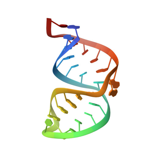
Structure of the A site of Escherichia coli 16S ribosomal RNA complexed with an aminoglycoside antibiotic.

Fourmy, D.Recht, M.I.Blanchard, S.C.Puglisi, J.D.

(1996) Science 274: 1367-1371

  • DOI: https://doi.org/10.1126/science.274.5291.1367
  • Primary Citation Related Structures: 
    1PBR

  • PubMed Abstract: 

    Aminoglycoside antibiotics that bind to 30S ribosomal A-site RNA cause misreading of the genetic code and inhibit translocation. The aminoglycoside antibiotic paromomycin binds specifically to an RNA oligonucleotide that contains the 30S subunit A site, and the solution structure of the RNA-paromomycin complex was determined by nuclear magnetic resonance spectroscopy. The antibiotic binds in the major groove of the model A-site RNA within a pocket created by an A-A base pair and a single bulged adenine. Specific interactions occur between aminoglycoside chemical groups important for antibiotic activity and conserved nucleotides in the RNA. The structure explains binding of diverse aminoglycosides to the ribosome, their specific activity against prokaryotic organisms, and various resistance mechanisms, and provides insight into ribosome function.


  • Organizational Affiliation
    • Department of Chemistry and Biochemistry, Center for Molecular Biology of RNA, University of California, Santa Cruz, CA 95064, USA.

Macromolecule Content 

  • Total Structure Weight: 9.31 kDa 
  • Atom Count: 617 
  • Modeled Residue Count: 27 
  • Deposited Residue Count: 27 
  • Unique nucleic acid chains: 1

Macromolecules

Find similar nucleic acids by:  (by identity cutoff) 
Entity ID: 1
MoleculeChains LengthOrganismImage
16S RIBOSOMAL RNA27N/A
Sequence Annotations
Expand
Reference Sequence

Oligosaccharides

Help  
Entity ID: 2
MoleculeChains Length2D Diagram GlycosylationD Interactions
2,6-diamino-2,6-dideoxy-beta-L-idopyranose-(1-3)-beta-D-ribofuranose
B
2N/A
Glycosylation Resources
GlyTouCan: G38023GV
GlyCosmos: G38023GV

Small Molecules

Ligands 2 Unique
IDChains Name / Formula / InChI Key2D Diagram3D Interactions
PA1

Query on PA1


Download:Ideal Coordinates CCD File
C [auth A]2-amino-2-deoxy-alpha-D-glucopyranose
C6 H13 N O5
MSWZFWKMSRAUBD-UKFBFLRUSA-N
CYY

Query on CYY


Download:Ideal Coordinates CCD File
D [auth A]2-DEOXYSTREPTAMINE
C6 H14 N2 O3
DTFAJAKTSMLKAT-JDCCYXBGSA-N

Experimental Data & Validation

Experimental Data

  • Method: SOLUTION NMR
  • Conformers Submitted: 

Structure Validation

View Full Validation Report


Entry History 

Deposition Data

Revision History  (Full details and data files)

  • Version 1.0: 1997-09-17
    Type: Initial release
  • Version 1.1: 2008-03-24
    Changes: Version format compliance
  • Version 1.2: 2011-07-13
    Changes: Version format compliance
  • Version 1.3: 2013-01-16
    Changes: Non-polymer description
  • Version 2.0: 2020-07-29
    Type: Remediation
    Reason: Carbohydrate remediation
    Changes: Atomic model, Data collection, Derived calculations, Other, Structure summary
  • Version 2.1: 2024-05-01
    Changes: Data collection, Database references, Structure summary