1PAH | pdb_00001pah

HUMAN PHENYLALANINE HYDROXYLASE DIMER, RESIDUES 117-424


Experimental Data Snapshot

  • Method: X-RAY DIFFRACTION
  • Resolution: 2.00 Å
  • R-Value Free: 
    0.218 (Depositor) 
  • R-Value Work: 
    0.176 (Depositor) 
  • R-Value Observed: 
    0.176 (Depositor) 

Starting Model: experimental
View more details

wwPDB Validation 3D Report Full Report

Validation slider image for 1PAH

This is version 1.4 of the entry. See complete history

Literature

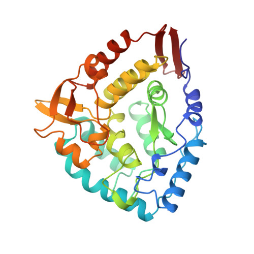
Crystal structure of the catalytic domain of human phenylalanine hydroxylase reveals the structural basis for phenylketonuria.

Erlandsen, H.Fusetti, F.Martinez, A.Hough, E.Flatmark, T.Stevens, R.C.

(1997) Nat Struct Biol 4: 995-1000

  • DOI: https://doi.org/10.1038/nsb1297-995
  • Primary Citation Related Structures: 
    1PAH

  • PubMed Abstract: 

    The 2.0 A crystal structure of the catalytic domain of human phenylalanine hydroxylase reveals a fold similar to that of tyrosine hydroxylase. It provides the first structural view of where mutations occur and a rationale to explain molecular mechanisms of the enzymatic phenotypes in the autosomal recessive disorder phenylketoneuria.

Macromolecule Content 

  • Total Structure Weight: 35.76 kDa 
  • Atom Count: 2,584 
  • Modeled Residue Count: 308 
  • Deposited Residue Count: 308 
  • Unique protein chains: 1

Macromolecules

Find similar proteins by:|  3D Structure
Entity ID: 1
MoleculeChains  Sequence LengthOrganismDetailsImage
PHENYLALANINE HYDROXYLASE308Homo sapiensMutation(s): 0 
EC: 1.14.16.1
UniProt & NIH Common Fund Data Resources
Find proteins for P00439 (Homo sapiens)
Explore P00439 
Go to UniProtKB:  P00439
PHAROS:  P00439
GTEx:  ENSG00000171759 
Entity Groups
Sequence Clusters30% Identity50% Identity70% Identity90% Identity95% Identity100% Identity
UniProt GroupP00439
Sequence Annotations
Expand
Reference Sequence

Small Molecules

Ligands 1 Unique
IDChains Name / Formula / InChI Key2D Diagram3D Interactions
FE

Query on FE


Download:Ideal Coordinates CCD File
B [auth A]FE (III) ION
Fe
VTLYFUHAOXGGBS-UHFFFAOYSA-N

Experimental Data & Validation

Experimental Data

  • Method: X-RAY DIFFRACTION
  • Resolution: 2.00 Å
  • R-Value Free:  0.218 (Depositor) 
  • R-Value Work:  0.176 (Depositor) 
  • R-Value Observed: 0.176 (Depositor) 
Space Group: C 2 2 21
Unit Cell:
Length ( Å )Angle ( ˚ )
a = 66.6α = 90
b = 108.4β = 90
c = 125.7γ = 90
Software Package:
Software NamePurpose
X-PLORmodel building
X-PLORrefinement
DENZOdata reduction
SCALEPACKdata scaling
X-PLORphasing

Structure Validation

View Full Validation Report


Entry History 

Deposition Data

Revision History  (Full details and data files)

  • Version 1.0: 1999-01-13
    Type: Initial release
  • Version 1.1: 2008-03-24
    Changes: Version format compliance
  • Version 1.2: 2011-07-13
    Changes: Version format compliance
  • Version 1.3: 2023-08-09
    Changes: Database references, Derived calculations, Other, Refinement description
  • Version 1.4: 2024-05-22
    Changes: Data collection