1P9L | pdb_00001p9l

Structure of M. tuberculosis dihydrodipicolinate reductase in complex with NADH and 2,6 PDC


Experimental Data Snapshot

  • Method: X-RAY DIFFRACTION
  • Resolution: 2.30 Å
  • R-Value Free: 
    0.250 (Depositor), 0.197 (DCC) 
  • R-Value Work: 
    0.193 (Depositor), 0.195 (DCC) 
  • R-Value Observed: 
    0.197 (Depositor) 

Starting Model: experimental
View more details

wwPDB Validation 3D Report Full Report

Validation slider image for 1P9L

Ligand Structure Quality Assessment 


This is version 1.4 of the entry. See complete history

Literature

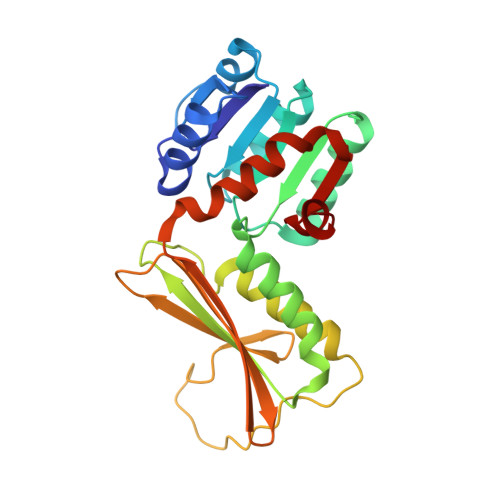
The three-dimensional structures of the Mycobacterium tuberculosis dihydrodipicolinate reductase-NADH-2,6-PDC and -NADPH-2,6-PDC complexes. Structural and mutagenic analysis of relaxed nucleotide specificity.

Cirilli, M.Zheng, R.Scapin, G.Blanchard, J.S.

(2003) Biochemistry 42: 10644-10650

  • DOI: https://doi.org/10.1021/bi030044v
  • Primary Citation Related Structures: 
    1C3V, 1P9L

  • PubMed Abstract: 

    Dihydrodipicolinate reductase (DHPR) catalyzes the reduced pyridine nucleotide-dependent reduction of the alpha,beta-unsaturated cyclic imine, dihydrodipicolinate, to generate tetrahydrodipicolinate. This enzyme catalyzes the second step in the bacterial biosynthetic pathway that generates meso-diaminopimelate, a component of bacterial cell walls, and the amino acid L-lysine. The Mycobacterium tuberculosis dapB-encoded DHPR has been cloned, expressed, purified, and crystallized in two ternary complexes with NADH or NADPH and the inhibitor 2,6-pyridinedicarboxylate (2,6-PDC). The structures have been solved using molecular replacement strategies, and the DHPR-NADH-2,6-PDC and DHPR-NADPH-2,6-PDC complexes have been refined against data to 2.3 and 2.5 A, respectively. The M. tuberculosis DHPR is a tetramer of identical subunits, with each subunit composed of two domains connected by two flexible hinge regions. The N-terminal domain binds pyridine nucleotide, while the C-terminal domain is involved in both tetramer formation and substrate/inhibitor binding. The M. tuberculosis DHPR uses NADH and NADPH with nearly equal efficiency based on V/K values. To probe the nature of this substrate specificity, we have generated two mutants, K9A and K11A, residues that are close to the 2'-phosphate of NADPH. These two mutants exhibit decreased specificity for NADPH by factors of 6- and 30-fold, respectively, but the K11A mutant exhibits 270% of WT activity using NADH. The highly conserved structure of the nucleotide fold may permit other enzyme's nucleotide specificity to be altered using similar mutagenic strategies.


  • Organizational Affiliation
    • Department of Biochemistry, Albert Einstein College of Medicine, 1300 Morris Park Avenue, Bronx, New York 10461, USA.

Macromolecule Content 

  • Total Structure Weight: 53.45 kDa 
  • Atom Count: 3,866 
  • Modeled Residue Count: 490 
  • Deposited Residue Count: 490 
  • Unique protein chains: 1

Macromolecules

Find similar proteins by:|  3D Structure
Entity ID: 1
MoleculeChains  Sequence LengthOrganismDetailsImage
dihydrodipicolinate reductase
A, B
245Mycobacterium tuberculosisMutation(s): 0 
Gene Names: dapB
EC: 1.3.1.26 (PDB Primary Data), 1.17.1.8 (UniProt)
UniProt
Find proteins for P9WP23 (Mycobacterium tuberculosis (strain ATCC 25618 / H37Rv))
Explore P9WP23 
Go to UniProtKB:  P9WP23
Entity Groups
Sequence Clusters30% Identity50% Identity70% Identity90% Identity95% Identity100% Identity
UniProt GroupP9WP23
Sequence Annotations
Expand
Reference Sequence

Experimental Data & Validation

Experimental Data

  • Method: X-RAY DIFFRACTION
  • Resolution: 2.30 Å
  • R-Value Free:  0.250 (Depositor), 0.197 (DCC) 
  • R-Value Work:  0.193 (Depositor), 0.195 (DCC) 
  • R-Value Observed: 0.197 (Depositor) 
Space Group: I 21 21 21
Unit Cell:
Length ( Å )Angle ( ˚ )
a = 119.29α = 90
b = 117.28β = 90
c = 78.54γ = 90
Software Package:
Software NamePurpose
Sadiedata collection
SAINTdata reduction
AMoREphasing
X-PLORrefinement
SADIEdata reduction
SAINTdata scaling

Structure Validation

View Full Validation Report


Ligand Structure Quality Assessment 


Entry History 

Deposition Data

Revision History  (Full details and data files)

  • Version 1.0: 2003-08-26
    Type: Initial release
  • Version 1.1: 2008-04-29
    Changes: Version format compliance
  • Version 1.2: 2011-07-13
    Changes: Derived calculations, Version format compliance
  • Version 1.3: 2013-11-06
    Changes: Non-polymer description
  • Version 1.4: 2023-08-16
    Changes: Data collection, Database references, Derived calculations, Refinement description