1P8D | pdb_00001p8d

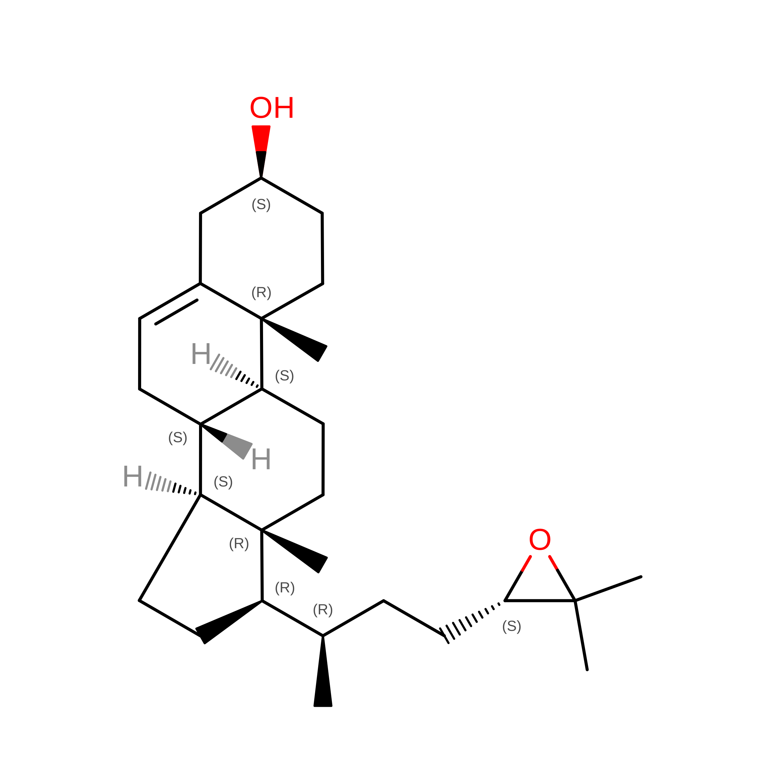
X-Ray Crystal Structure of LXR Ligand Binding Domain with 24(S),25-epoxycholesterol


Experimental Data Snapshot

  • Method: X-RAY DIFFRACTION
  • Resolution: 2.80 Å
  • R-Value Free: 
    0.277 (Depositor) 
  • R-Value Work: 
    0.214 (Depositor) 
  • R-Value Observed: 
    0.214 (Depositor) 

Starting Model: experimental
View more details

wwPDB Validation 3D Report Full Report

Validation slider image for 1P8D

This is version 1.6 of the entry. See complete history. 

Literature

X-ray crystal structure of the liver X receptor beta ligand binding domain: regulation by a histidine-tryptophan switch.

Williams, S., Bledsoe, R.K., Collins, J.L., Boggs, S., Lambert, M.H., Miller, A.B., Moore, J., McKee, D.D., Moore, L., Nichols, J., Parks, D., Watson, M., Wisely, B., Willson, T.M.

(2003) J Biol Chem 278: 27138-27143

  • DOI: https://doi.org/10.1074/jbc.M302260200
  • Primary Citation Related Structures: 
    1P8D

  • PubMed Abstract: 

    The x-ray crystal structures of the human liver X receptor beta ligand binding domain complexed to sterol and nonsterol agonists revealed a perpendicular histidinetryptophan switch that holds the receptor in its active conformation. Hydrogen bonding interactions with the ligand act to position the His-435 imidazole ring against the Trp-457 indole ring, allowing an electrostatic interaction that holds the AF2 helix in the active position. The neutral oxysterol 24(S),25-epoxycholesterol accepts a hydrogen bond from His-435 that positions the imidazole ring of the histidine above the pyrrole ring of the tryptophan. In contrast, the acidic T0901317 hydroxyl group makes a shorter hydrogen bond with His-435 that pulls the imidazole over the electron-rich benzene ring of the tryptophan, possibly strengthening the electrostatic interaction. Point mutagenesis of Trp-457 supports the observation that the ligand-histidine-tryptophan coupling is different between the two ligands. The lipophilic liver X receptor ligand-binding pocket is larger than the corresponding steroid hormone receptors, which allows T0901317 to adopt two distinct conformations. These results provide a molecular basis for liver X receptor activation by a wide range of endogenous neutral and acidic ligands.


  • Organizational Affiliation: 
    • GlaxoSmithKline, Discovery Research, Research Triangle Park, NC 27709, USA. shawn.p.williams@gsk.com

Macromolecule Content 

  • Total Structure Weight: 63.88 kDa 
  • Atom Count: 4,273 
  • Modeled Residue Count: 508 
  • Deposited Residue Count: 550 
  • Unique protein chains: 2

Macromolecules

Find similar proteins by:|  3D Structure
Entity ID: 1
MoleculeChains  Sequence LengthOrganismDetailsImage
Oxysterols receptor LXR-beta
A, B
250Homo sapiensMutation(s): 0 
Gene Names: NR1H2 OR LXRB OR UNR OR NER
UniProt & NIH Common Fund Data Resources
Find proteins for P55055 (Homo sapiens)
Explore P55055 
Go to UniProtKB:  P55055
PHAROS:  P55055
GTEx:  ENSG00000131408 
Entity Groups
Sequence Clusters30% Identity50% Identity70% Identity90% Identity95% Identity100% Identity
UniProt GroupP55055
Sequence Annotations
Expand
Reference Sequence
Find similar proteins by:  Sequence   |   3D Structure  
Entity ID: 2
MoleculeChains  Sequence LengthOrganismDetailsImage
nuclear receptor coactivator 1 isoform 3
C, D
25N/AMutation(s): 0 
EC: 2.3.1.48
UniProt & NIH Common Fund Data Resources
Find proteins for Q15788 (Homo sapiens)
Explore Q15788 
Go to UniProtKB:  Q15788
PHAROS:  Q15788
GTEx:  ENSG00000084676 
Entity Groups
Sequence Clusters30% Identity50% Identity70% Identity90% Identity95% Identity100% Identity
UniProt GroupQ15788
Sequence Annotations
Expand
Reference Sequence

Small Molecules

Ligands 1 Unique
IDChains Name / Formula / InChI Key2D Diagram3D Interactions
CO1

Query on CO1


Download:Ideal Coordinates CCD File
E [auth A],
F [auth B]
17-[3-(3,3-DIMETHYL-OXIRANYL)-1-METHYL-PROPYL]-10,13-DIMETHYL-2,3,4,7,8,9,10,11,12,13,14,15,16,17-TETRADECAHYDRO-1H-CYC LOPENTA[A]PHENANTHREN-3-OL
C27 H44 O2
OSENKJZWYQXHBN-XVYZBDJZSA-N

Experimental Data & Validation

Experimental Data

  • Method: X-RAY DIFFRACTION
  • Resolution: 2.80 Å
  • R-Value Free:  0.277 (Depositor) 
  • R-Value Work:  0.214 (Depositor) 
  • R-Value Observed: 0.214 (Depositor) 
Space Group: C 2 2 21
Unit Cell:
Length ( Å )Angle ( ˚ )
a = 71.166α = 90
b = 120.012β = 90
c = 147.56γ = 90
Software Package:
Software NamePurpose
MAR345data collection
SCALEPACKdata scaling
AMoREphasing
CNSrefinement

Structure Validation

View Full Validation Report


Entry History 

Revision History  (Full details and data files)

  • Version 1.0: 2003-07-08
    Type: Initial release
  • Version 1.1: 2008-04-29
    Changes: Version format compliance
  • Version 1.2: 2011-07-13
    Changes: Version format compliance
  • Version 1.3: 2011-11-16
    Changes: Atomic model
  • Version 1.4: 2017-10-11
    Changes: Refinement description
  • Version 1.5: 2024-02-14
    Changes: Data collection, Database references, Derived calculations, Structure summary
  • Version 1.6: 2024-04-03
    Changes: Refinement description