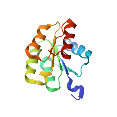
Solution Structures of the Inactive and BeF(3)-activated Response Regulator CheY2
Riepl, H., Scharf, B., Schmitt, R., Kalbitzer, H.R., Maurer, T.(2004) J Biological Chem 338: 287-297
- PubMed: 15066432 Search on PubMed
- DOI: https://doi.org/10.1016/j.jmb.2004.02.054
- Primary Citation Related Structures:
1P6Q, 1P6U - PubMed Abstract:
The chemotactic signalling chain to the flagellar motor of Sinorhizobium meliloti features a new type of response regulator, CheY2. CheY2 activated by phosphorylation (CheY2-P) controls the rotary speed of the flagellar motor (instead of reversing the sense of rotation), and it is efficiently dephosphorylated by phospho-retrotransfer to the cognate kinase, CheA. Here, we report the NMR solution structures of the Mg(2+)-complex of inactive CheY2, and of activated CheY2-BeF(3), a stable analogue of CheY2-P, to an overall root mean square deviation of 0.042 nm and 0.027 nm, respectively. The 14 kDa CheY2 protein exhibits a characteristic open (alpha/beta)(5) conformation. Modification of CheY2 by BeF(3)(-) leads to large conformational changes of the protein, which are in the limits of error identical with those observed by phosphorylation of the active-centre residue Asp58. In BeF(3)-activated CheY2, the position of Thr88-OH favours the formation of a hydrogen bond with the active site, Asp58-BeF(3), similar to BeF(3)-activated CheY from Escherichia coli. In contrast to E.coli, this reorientation is not involved in a Tyr-Thr-coupling mechanism, that propagates the signal from the incoming phosphoryl group to the C-terminally located FliM-binding surface. Rather, a rearrangement of the Phe59 side-chain to interact with Ile86-Leu95-Val96 along with a displacement of alpha4 towards beta5 is stabilised in S.meliloti. The resulting, activation-induced, compact alpha4-beta5-alpha5 surface forms a unique binding domain suited for specific interaction with and signalling to a rotary motor that requires a gradual speed control. We propose that these new features of response regulator activation, compared to other two-component systems, are the key for the observed unique phosphorylation, dephosphorylation and motor control mechanisms in S.meliloti.
- Lehrstuhl für Genetik, Universität Regensburg, D-93040 Regensburg, Germany.
Organizational Affiliation: