Structural and Biochemical Characterization of Toad Liver Basic Fatty Acid-Binding Protein
Di Pietro, S.M., Corsico, B., Perduca, M., Monaco, H.L., Santome, J.A.(2003) Biochemistry 42: 8192-8203
- PubMed: 12846568 Search on PubMed
- DOI: https://doi.org/10.1021/bi034213n
- Primary Citation Related Structures:
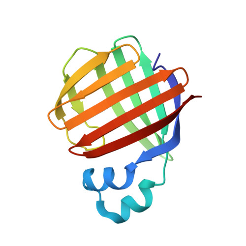
1P6P - PubMed Abstract:
Two paralogous groups of fatty acid-binding proteins (FABPs) have been described in vertebrate liver: liver FABP (L-FABP) type, extensively characterized in mammals, and liver basic FABP (Lb-FABP) found in fish, amphibians, reptiles, and birds. We describe here the toad Lb-FABP complete amino acid sequence, its X-ray structure to 2.5 A resolution, ligand-binding properties, and mechanism of fatty acid transfer to phospholipid membranes. Alignment of the amino acid sequence of toad Lb-FABP with known L-FABPs and Lb-FABPs shows that it is more closely related to the other Lb-FABPs. Toad Lb-FABP conserves the 12 characteristic residues present in all Lb-FABPs and absent in L-FABPs and presents the canonical fold characteristic of all the members of this protein family. Eight out of the 12 conserved residues point to the lipid-binding cavity of the molecule. In contrast, most of the 25 L-FABP conserved residues are in clusters on the surface of the molecule. The helix-turn-helix motif shows both a negative and positive electrostatic potential surface as in rat L-FABP, and in contrast with the other FABP types. The mechanism of anthroyloxy-labeled fatty acids transfer from Lb-FABP to phospholipid membranes occurs by a diffusion-mediated process, as previously shown for L-FABP, but the rate of transfer is 1 order of magnitude faster. Toad Lb-FABP can bind two cis-parinaric acid molecules but only one trans-parinaric acid molecule while L-FABP binds two molecules of both parinaric acid isomers. Although toad Lb-FABP shares with L-FABP a broad ligand-binding specificity, the relative affinity is different.
- Instituto de Química y Fisicoquímica Biológicas (IQUIFIB)-CONICET-UBA, Facultad de Farmacia y Bioquímica, UBA, Junín 956, Buenos Aires 1113, Argentina. sdipietro@mednet.ucla.edu
Organizational Affiliation: