1P4T

Crystal structure of Neisserial surface protein A (NspA)


Experimental Data Snapshot

  • Method: X-RAY DIFFRACTION
  • Resolution: 2.55 Å
  • R-Value Free: 
    0.258 (Depositor), 0.260 (DCC) 
  • R-Value Work: 
    0.225 (Depositor), 0.220 (DCC) 
  • R-Value Observed: 
    0.227 (Depositor) 

wwPDB Validation   3D Report Full Report


Ligand Structure Quality Assessment 

Created with Raphaël 2.3.0Worse 01 BetterLigand structure goodness of fit to experimental dataBest fitted CXEClick on this verticalbar to view details

This is version 2.0 of the entry. See complete history


Literature

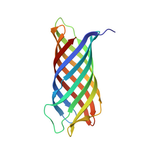
Crystal structure of Neisserial Surface Protein A (NspA), a conserved outer membrane protein with vaccine potential

Vandeputte-Rutten, L.Bos, M.P.Tommassen, J.Gros, P.

(2003) J Biol Chem 278: 24825-24830

  • DOI: https://doi.org/10.1074/jbc.M302803200
  • Primary Citation of Related Structures:  
    1P4T

  • PubMed Abstract: 

    The neisserial surface protein A (NspA) from Neisseria meningitidis is a promising vaccine candidate because it is highly conserved among meningococcal strains and induces bactericidal antibodies. NspA is a homolog of the Opa proteins, which mediate adhesion to host cells. Here, we present the crystal structure of NspA, determined to 2.55-A resolution. NspA forms an eight-stranded antiparallel beta-barrel. The four loops at the extracellular side of the NspA molecule form a long cleft, which contains mainly hydrophobic residues and harbors a detergent molecule, suggesting that the protein might function in the binding of hydrophobic ligands, such as lipids. In addition, the structure provides a starting point for structure-based vaccine design.


  • Organizational Affiliation

    Department of Crystal and Structural Chemistry, Bijvoet Center for Biomolecular Research, Utrecht, The Netherlands.


Macromolecules
Find similar proteins by:  (by identity cutoff)  |  3D Structure
Entity ID: 1
MoleculeChains Sequence LengthOrganismDetailsImage
outer membrane protein NspA155Neisseria meningitidisMutation(s): 0 
Gene Names: nspa
Membrane Entity: Yes 
UniProt
Find proteins for Q9RP17 (Neisseria meningitidis)
Explore Q9RP17 
Go to UniProtKB:  Q9RP17
Entity Groups  
Sequence Clusters30% Identity50% Identity70% Identity90% Identity95% Identity100% Identity
UniProt GroupQ9RP17
Sequence Annotations
Expand
  • Reference Sequence
Small Molecules
Ligands 3 Unique
IDChains Name / Formula / InChI Key2D Diagram3D Interactions
CXE
Query on CXE

Download Ideal Coordinates CCD File 
C [auth A],
D [auth A],
E [auth A],
F [auth A],
G [auth A]
PENTAETHYLENE GLYCOL MONODECYL ETHER
C20 H42 O6
QAXPOSPGRHYIHE-UHFFFAOYSA-N
SO4
Query on SO4

Download Ideal Coordinates CCD File 
B [auth A]SULFATE ION
O4 S
QAOWNCQODCNURD-UHFFFAOYSA-L
ETA
Query on ETA

Download Ideal Coordinates CCD File 
H [auth A]ETHANOLAMINE
C2 H7 N O
HZAXFHJVJLSVMW-UHFFFAOYSA-N
Experimental Data & Validation

Experimental Data

  • Method: X-RAY DIFFRACTION
  • Resolution: 2.55 Å
  • R-Value Free:  0.258 (Depositor), 0.260 (DCC) 
  • R-Value Work:  0.225 (Depositor), 0.220 (DCC) 
  • R-Value Observed: 0.227 (Depositor) 
Space Group: H 3 2
Unit Cell:
Length ( Å )Angle ( ˚ )
a = 97.372α = 90
b = 97.372β = 90
c = 171.939γ = 120
Software Package:
Software NamePurpose
REFMACrefinement
DENZOdata reduction
SCALEPACKdata scaling
MLPHAREphasing

Structure Validation

View Full Validation Report


Ligand Structure Quality Assessment 

Created with Raphaël 2.3.0Worse 01 BetterLigand structure goodness of fit to experimental dataBest fitted CXEClick on this verticalbar to view details

Entry History 

Deposition Data

Revision History  (Full details and data files)

  • Version 1.0: 2003-07-22
    Type: Initial release
  • Version 1.1: 2008-04-29
    Changes: Version format compliance
  • Version 1.2: 2011-07-13
    Changes: Advisory, Derived calculations, Version format compliance
  • Version 2.0: 2023-11-15
    Changes: Atomic model, Data collection, Database references, Derived calculations