1OXA | pdb_00001oxa

CYTOCHROME P450 (DONOR:O2 OXIDOREDUCTASE)


Experimental Data Snapshot

  • Method: X-RAY DIFFRACTION
  • Resolution: 2.10 Å
  • R-Value Work: 
    0.196 (Depositor) 
  • R-Value Observed: 
    0.196 (Depositor) 

wwPDB Validation 3D Report Full Report

Validation slider image for 1OXA

This is version 1.3 of the entry. See complete history

Literature

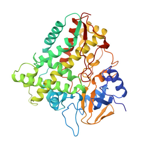
Structure of cytochrome P450eryF involved in erythromycin biosynthesis.

Cupp-Vickery, J.R.Poulos, T.L.

(1995) Nat Struct Biol 2: 144-153

  • DOI: https://doi.org/10.1038/nsb0295-144
  • Primary Citation Related Structures: 
    1OXA

  • PubMed Abstract: 

    Cytochrome P450eryF catalyzes the 6S-hydroxylation of 6-deoxyerythronolide B, the initial reaction in a multistep pathway to convert 6-deoxyerythronolide B into the antibiotic, erythromycin. The overall structure of P450eryF is similar to that of P450cam but differs in the exact positioning of several alpha-helices. The largest difference occurs in the B' helix and results in the enlargement of the substrate-binding pocket of P450eryF. The substrate is positioned with the macrolide ring perpendicular to the haem plane and contacts seven hydrophobic residues and three solvent molecules. The substrate participates in a network of hydrogen bonds that may provide a proton shuttle pathway in the oxygen cleavage reaction.


  • Organizational Affiliation
    • Department of Molecular Biology and Biochemistry, University of California, Irvine 92717, USA.

Macromolecule Content 

  • Total Structure Weight: 45.52 kDa 
  • Atom Count: 3,450 
  • Modeled Residue Count: 403 
  • Deposited Residue Count: 403 
  • Unique protein chains: 1

Macromolecules

Find similar proteins by:|  3D Structure
Entity ID: 1
MoleculeChains  Sequence LengthOrganismDetailsImage
CYTOCHROME P450 ERYF403Saccharopolyspora erythraeaMutation(s): 0 
EC: 1.14.15.35
UniProt
Find proteins for Q00441 (Saccharopolyspora erythraea (strain ATCC 11635 / DSM 40517 / JCM 4748 / NBRC 13426 / NCIMB 8594 / NRRL 2338))
Explore Q00441 
Go to UniProtKB:  Q00441
Entity Groups
Sequence Clusters30% Identity50% Identity70% Identity90% Identity95% Identity100% Identity
UniProt GroupQ00441
Sequence Annotations
Expand
Reference Sequence

Small Molecules

Ligands 2 Unique
IDChains Name / Formula / InChI Key2D Diagram3D Interactions
HEM

Query on HEM


Download:Ideal Coordinates CCD File
B [auth A]PROTOPORPHYRIN IX CONTAINING FE
C34 H32 Fe N4 O4
KABFMIBPWCXCRK-RGGAHWMASA-L
DEB

Query on DEB


Download:Ideal Coordinates CCD File
C [auth A]6-DEOXYERYTHRONOLIDE B
C21 H38 O6
HQZOLNNEQAKEHT-IBBGRPSASA-N

Experimental Data & Validation

Experimental Data

  • Method: X-RAY DIFFRACTION
  • Resolution: 2.10 Å
  • R-Value Work:  0.196 (Depositor) 
  • R-Value Observed: 0.196 (Depositor) 
Space Group: P 21 21 21
Unit Cell:
Length ( Å )Angle ( ˚ )
a = 54.16α = 90
b = 79.67β = 90
c = 99.48γ = 90
Software Package:
Software NamePurpose
X-PLORmodel building
X-PLORrefinement
XENGENdata reduction
SDMSdata reduction
X-PLORphasing

Structure Validation

View Full Validation Report


Entry History 

Deposition Data

Revision History  (Full details and data files)

  • Version 1.0: 1995-12-07
    Type: Initial release
  • Version 1.1: 2008-03-24
    Changes: Version format compliance
  • Version 1.2: 2011-07-13
    Changes: Version format compliance
  • Version 1.3: 2024-02-14
    Changes: Data collection, Database references, Derived calculations, Other