1OSN | pdb_00001osn

Crystal structure of Varicella zoster virus thymidine kinase in complex with BVDU-MP and ADP


Experimental Data Snapshot

  • Method: X-RAY DIFFRACTION
  • Resolution: 3.20 Å
  • R-Value Free: 
    0.268 (Depositor), 0.248 (DCC) 
  • R-Value Work: 
    0.235 (Depositor), 0.215 (DCC) 

Starting Model: experimental
View more details

wwPDB Validation 3D Report Full Report

Validation slider image for 1OSN

Ligand Structure Quality Assessment 


This is version 1.3 of the entry. See complete history

Literature

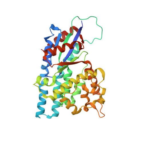
Crystal structure of varicella zoster virus thymidine kinase

Bird, L.E.Ren, J.Wright, A.Leslie, K.D.Degreve, B.Balzarini, J.Stammers, D.K.

(2003) J Biological Chem 278: 24680-24687

  • DOI: https://doi.org/10.1074/jbc.M302025200
  • Primary Citation Related Structures: 
    1OSN

  • PubMed Abstract: 

    Herpes virus thymidine kinases are responsible for the activation of nucleoside antiviral drugs including (E)-5-(2-bromovinyl)-2'-deoxyuridine. Such viral thymidine kinases (tk), beside having a broader substrate specificity compared with host cell enzymes, also show significant variation in nucleoside phosphorylation among themselves. We have determined the crystal structure of Varicella zoster virus (VZV, human herpes virus 3) thymidine kinase complexed with (E)-5-(2-bromovinyl)-2'-deoxyuridine 5'-monophosphate and ADP. Differences in the conformation of a loop region (residues 55-61) and the position of two alpha-helices at the subunit interface of VZV-tk compared with the herpes simplex virus type 1 (human herpes virus 1) enzyme give rise to changes in the positioning of residues such as tyrosine 66 and glutamine 90, which hydrogen bond to the substrate in the active site. Such changes in combination with the substitution in VZV-tk of two phenylalanine residues (in place of a tyrosine and methionine), which sandwich the substrate pyrimidine ring, cause an alteration in the positioning of the base. The interaction of the (E)-5-(2-bromovinyl)-2'-deoxyuridine deoxyribose ring with the protein is altered by substitution of tyrosine 21 and phenylalanine 139 (analagous to herpes simplex virus type 1 histidine 58 and tyrosine 172), which may explain some of the differences in nucleoside sugar selectivity between both enzymes. The altered active site architecture may also account for the differences in the substrate activity of ganciclovir for the two thymidine kinases. These data should be of use in the design of novel antiherpes and antitumor drugs.


  • Organizational Affiliation
    • Division of Structural Biology, The Wellcome Trust Centre for Human Genetics, Henry Wellcome Building of Genomic Medicine, University of Oxford, Roosevelt Drive, Headington, United Kingdom.

Macromolecule Content 

  • Total Structure Weight: 154.88 kDa 
  • Atom Count: 10,038 
  • Modeled Residue Count: 1,268 
  • Deposited Residue Count: 1,364 
  • Unique protein chains: 1

Macromolecules

Find similar proteins by:|  3D Structure
Entity ID: 1
MoleculeChains  Sequence LengthOrganismDetailsImage
Thymidine kinase
A, B, C, D
341Human alphaherpesvirus 3Mutation(s): 0 
EC: 2.7.1.21
UniProt
Find proteins for P0C0E6 (Varicella-zoster virus (strain Oka vaccine))
Explore P0C0E6 
Go to UniProtKB:  P0C0E6
Entity Groups
Sequence Clusters30% Identity50% Identity70% Identity90% Identity95% Identity100% Identity
UniProt GroupP0C0E6
Sequence Annotations
Expand
Reference Sequence

Small Molecules

Ligands 2 Unique
IDChains Name / Formula / InChI Key2D Diagram3D Interactions
ADP

Query on ADP


Download:Ideal Coordinates CCD File
E [auth A],
G [auth B],
I [auth C],
K [auth D]
ADENOSINE-5'-DIPHOSPHATE
C10 H15 N5 O10 P2
XTWYTFMLZFPYCI-KQYNXXCUSA-N
BVP

Query on BVP


Download:Ideal Coordinates CCD File
F [auth A],
H [auth B],
J [auth C],
L [auth D]
(E)-5-(2-BROMOVINYL)-2'-DEOXYURIDINE-5'-MONOPHOSPHATE
C11 H14 Br N2 O8 P
LKWCVKAHHUJPQO-PIXDULNESA-N

Experimental Data & Validation

Experimental Data

  • Method: X-RAY DIFFRACTION
  • Resolution: 3.20 Å
  • R-Value Free:  0.268 (Depositor), 0.248 (DCC) 
  • R-Value Work:  0.235 (Depositor), 0.215 (DCC) 
Space Group: P 1 21 1
Unit Cell:
Length ( Å )Angle ( ˚ )
a = 99.7α = 90
b = 54.2β = 94.8
c = 167.8γ = 90
Software Package:
Software NamePurpose
CNSrefinement
DENZOdata reduction
SCALEPACKdata scaling
CNSphasing

Structure Validation

View Full Validation Report


Ligand Structure Quality Assessment 


Entry History 

Deposition Data

Revision History  (Full details and data files)

  • Version 1.0: 2003-06-10
    Type: Initial release
  • Version 1.1: 2008-04-29
    Changes: Version format compliance
  • Version 1.2: 2011-07-13
    Changes: Version format compliance
  • Version 1.3: 2023-10-25
    Changes: Data collection, Database references, Derived calculations, Refinement description