1OS6 | pdb_00001os6

Cytochrome c7 (PpcA) from Geobacter sulfurreducens


Experimental Data Snapshot

  • Method: X-RAY DIFFRACTION
  • Resolution: 1.45 Å
  • R-Value Free: 
    0.227 (Depositor), 0.215 (DCC) 
  • R-Value Work: 
    0.182 (Depositor), 0.174 (DCC) 
  • R-Value Observed: 
    0.182 (Depositor) 

wwPDB Validation 3D Report Full Report

Validation slider image for 1OS6

Ligand Structure Quality Assessment 


This is version 2.1 of the entry. See complete history

Literature

Family of cytochrome c7-type proteins from Geobacter sulfurreducens: structure of one cytochrome c7 at 1.45 A resolution.

Pokkuluri, P.R.Londer, Y.Y.Duke, N.E.Long, W.C.Schiffer, M.

(2004) Biochemistry 43: 849-859

  • DOI: https://doi.org/10.1021/bi0301439
  • Primary Citation Related Structures: 
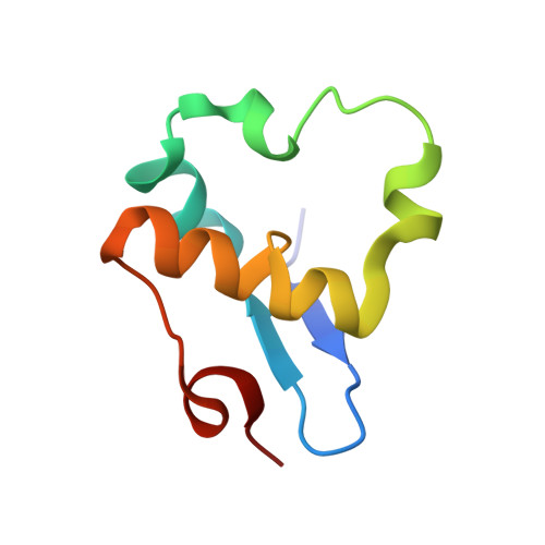
    1OS6

  • PubMed Abstract: 

    The structure of a cytochrome c(7) (PpcA) from Geobacter sulfurreducens was determined by X-ray diffraction at 1.45 A resolution; the R factor is 18.2%. The protein contains a three-heme core that is surrounded by 71 amino acid residues. An unusual feature of this cytochrome is that it has 17 lysine residues, but only nine hydrophobic residues that are larger than alanine. The details of the structure are described and compared with those of cytochrome c(7) from Desulfuromonas acetoxidans and with cytochromes c(3). The two cytochrome c(7) molecules have sequences that are 46% identical, but the arrangements of the hemes in the two structures differ; the rms deviation of all alpha-carbons is 2.5 A. These cytochromes can reduce various metal ions. The reduction site of the chromate ion in D. acetoxidans is occupied by a sulfate ion in the crystal structure of PpcA. We identified four additional homologues of cytochrome c(7) in the G. sulfurreducens genome and three polymers of c(7)-type domains. Of the polymers, two have four repeats and one has nine repeats. On the basis of sequence alignments, one of the hemes in each of the cytochrome c(7)-type domains does not have the bis-histidine coordination. The packing of the molecules in the crystal structure of PpcA suggests that the polymers have an elongated conformation and might form a "nanowire".


  • Organizational Affiliation
    • Biosciences Division, Argonne National Laboratory, Argonne, Illinois 60439, USA.

Macromolecule Content 

  • Total Structure Weight: 10.28 kDa 
  • Atom Count: 802 
  • Modeled Residue Count: 71 
  • Deposited Residue Count: 71 
  • Unique protein chains: 1

Macromolecules

Find similar proteins by:|  3D Structure
Entity ID: 1
MoleculeChains  Sequence LengthOrganismDetailsImage
PpcA71Geobacter sulfurreducensMutation(s): 0 
UniProt
Find proteins for Q8GGK7 (Geobacter sulfurreducens)
Explore Q8GGK7 
Go to UniProtKB:  Q8GGK7
Entity Groups
Sequence Clusters30% Identity50% Identity70% Identity90% Identity95% Identity100% Identity
UniProt GroupQ8GGK7
Sequence Annotations
Expand
Reference Sequence

Small Molecules

Ligands 3 Unique
IDChains Name / Formula / InChI Key2D Diagram3D Interactions
HEC

Query on HEC


Download:Ideal Coordinates CCD File
E [auth A],
F [auth A],
G [auth A]
HEME C
C34 H34 Fe N4 O4
HXQIYSLZKNYNMH-LJNAALQVSA-N
DXC

Query on DXC


Download:Ideal Coordinates CCD File
H [auth A](3ALPHA,5BETA,12ALPHA)-3,12-DIHYDROXYCHOLAN-24-OIC ACID
C24 H40 O4
KXGVEGMKQFWNSR-LLQZFEROSA-N
SO4

Query on SO4


Download:Ideal Coordinates CCD File
B [auth A],
C [auth A],
D [auth A]
SULFATE ION
O4 S
QAOWNCQODCNURD-UHFFFAOYSA-L

Experimental Data & Validation

Experimental Data

  • Method: X-RAY DIFFRACTION
  • Resolution: 1.45 Å
  • R-Value Free:  0.227 (Depositor), 0.215 (DCC) 
  • R-Value Work:  0.182 (Depositor), 0.174 (DCC) 
  • R-Value Observed: 0.182 (Depositor) 
Space Group: P 43 2 2
Unit Cell:
Length ( Å )Angle ( ˚ )
a = 32.4α = 90
b = 32.4β = 90
c = 178.4γ = 90
Software Package:
Software NamePurpose
SHELXmodel building
SHELXL-97refinement
HKL-2000data reduction
SCALEPACKdata scaling
CNSphasing

Structure Validation

View Full Validation Report


Ligand Structure Quality Assessment 


Entry History 

Deposition Data

Revision History  (Full details and data files)

  • Version 1.0: 2004-02-03
    Type: Initial release
  • Version 1.1: 2008-04-29
    Changes: Version format compliance
  • Version 1.2: 2011-07-13
    Changes: Derived calculations, Version format compliance
  • Version 2.0: 2021-03-03
    Changes: Advisory, Atomic model, Data collection, Derived calculations, Non-polymer description, Structure summary
  • Version 2.1: 2024-11-20
    Changes: Data collection, Database references, Structure summary