1ON3 | pdb_00001on3

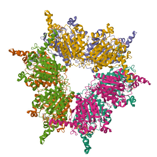
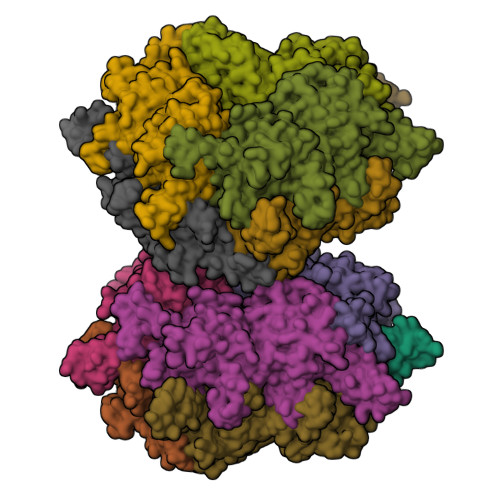
Transcarboxylase 12S crystal structure: hexamer assembly and substrate binding to a multienzyme core (with methylmalonyl-coenzyme a and methylmalonic acid bound)


Experimental Data Snapshot

  • Method: X-RAY DIFFRACTION
  • Resolution: 1.90 Å
  • R-Value Free: 
    0.213 (Depositor), 0.210 (DCC) 
  • R-Value Work: 
    0.168 (Depositor), 0.170 (DCC) 
  • R-Value Observed: 
    0.168 (Depositor) 

wwPDB Validation   3D Report Full Report


Ligand Structure Quality Assessment 

Created with Raphaël 2.3.0Worse 01 BetterLigand structure goodness of fit to experimental dataBest fitted MCAClick on this verticalbar to view details

This is version 1.3 of the entry. See complete history


Literature

Transcarboxylase 12S crystal structure: hexamer assembly and substrate binding to a multienzyme core

Hall, P.R.Wang, Y.-F.Rivera-Hainaj, R.E.Zheng, X.Pustai-Carey, M.Carey, P.R.Yee, V.C.

(2003) EMBO J 22: 2334-2347

  • DOI: https://doi.org/10.1093/emboj/cdg244
  • Primary Citation of Related Structures:  
    1ON3, 1ON9

  • PubMed Abstract: 

    Transcarboxylase from Propionibacterium shermanii is a 1.2 MDa multienzyme complex that couples two carboxylation reactions, transferring CO(2)(-) from methylmalonyl-CoA to pyruvate, yielding propionyl-CoA and oxaloacetate. The 1.9 A resolution crystal structure of the central 12S hexameric core, which catalyzes the first carboxylation reaction, has been solved bound to its substrate methylmalonyl-CoA. Overall, the structure reveals two stacked trimers related by 2-fold symmetry, and a domain duplication in the monomer. In the active site, the labile carboxylate group of methylmalonyl-CoA is stabilized by interaction with the N-termini of two alpha-helices. The 12S domains are structurally similar to the crotonase/isomerase superfamily, although only domain 1 of each 12S monomer binds ligand. The 12S reaction is similar to that of human propionyl-CoA carboxylase, whose beta-subunit has 50% sequence identity with 12S. A homology model of the propionyl-CoA carboxylase beta-subunit, based on this 12S crystal structure, provides new insight into the propionyl-CoA carboxylase mechanism, its oligomeric structure and the molecular basis of mutations responsible for enzyme deficiency in propionic acidemia.


  • Organizational Affiliation

    Department of Molecular Cardiology and Center for Structural Biology, Lerner Research Institute, Cleveland Clinic Foundation, OH 44195, USA.


Macromolecules
Find similar proteins by:  (by identity cutoff)  |  3D Structure
Entity ID: 1
MoleculeChains Sequence LengthOrganismDetailsImage
Methylmalonyl-CoA carboxyltransferase 12S subunit
A, B, C, D, E
523Propionibacterium freudenreichiiMutation(s): 0 
EC: 2.1.3.1
UniProt
Find proteins for Q8GBW6 (Propionibacterium freudenreichii subsp. shermanii)
Explore Q8GBW6 
Go to UniProtKB:  Q8GBW6
Entity Groups  
Sequence Clusters30% Identity50% Identity70% Identity90% Identity95% Identity100% Identity
UniProt GroupQ8GBW6
Sequence Annotations
Expand
  • Reference Sequence
Small Molecules
Ligands 4 Unique
IDChains Name / Formula / InChI Key2D Diagram3D Interactions
MCA
Query on MCA

Download Ideal Coordinates CCD File 
H [auth A]
L [auth B]
P [auth C]
R [auth D]
T [auth E]
METHYLMALONYL-COENZYME A
C25 H40 N7 O19 P3 S
MZFOKIKEPGUZEN-AGCMQPJKSA-N
MPD
Query on MPD

Download Ideal Coordinates CCD File 
J [auth A],
N [auth B],
X [auth F]
(4S)-2-METHYL-2,4-PENTANEDIOL
C6 H14 O2
SVTBMSDMJJWYQN-YFKPBYRVSA-N
DXX
Query on DXX

Download Ideal Coordinates CCD File 
I [auth A]
M [auth B]
Q [auth C]
S [auth D]
U [auth E]
METHYLMALONIC ACID
C4 H6 O4
ZIYVHBGGAOATLY-UHFFFAOYSA-N
CD
Query on CD

Download Ideal Coordinates CCD File 
G [auth A],
K [auth B],
O [auth C]
CADMIUM ION
Cd
WLZRMCYVCSSEQC-UHFFFAOYSA-N
Experimental Data & Validation

Experimental Data

  • Method: X-RAY DIFFRACTION
  • Resolution: 1.90 Å
  • R-Value Free:  0.213 (Depositor), 0.210 (DCC) 
  • R-Value Work:  0.168 (Depositor), 0.170 (DCC) 
  • R-Value Observed: 0.168 (Depositor) 
Space Group: C 1 2 1
Unit Cell:
Length ( Å )Angle ( ˚ )
a = 114.805α = 90
b = 200.708β = 102.43
c = 146.347γ = 90
Software Package:
Software NamePurpose
CNSrefinement
DENZOdata reduction
SCALEPACKdata scaling
SHARPphasing

Structure Validation

View Full Validation Report


Ligand Structure Quality Assessment 

Created with Raphaël 2.3.0Worse 01 BetterLigand structure goodness of fit to experimental dataBest fitted MCAClick on this verticalbar to view details

Entry History 

Deposition Data

Revision History  (Full details and data files)

  • Version 1.0: 2003-05-20
    Type: Initial release
  • Version 1.1: 2008-04-29
    Changes: Version format compliance
  • Version 1.2: 2011-07-13
    Changes: Version format compliance
  • Version 1.3: 2024-02-14
    Changes: Data collection, Database references, Derived calculations