1OEW | pdb_00001oew

ATOMIC RESOLUTION STRUCTURE OF NATIVE ENDOTHIAPEPSIN


Experimental Data Snapshot

  • Method: X-RAY DIFFRACTION
  • Resolution: 0.90 Å
  • R-Value Free: 
    0.147 (Depositor) 
  • R-Value Observed: 
    0.121 (Depositor) 

wwPDB Validation 3D Report Full Report

Validation slider image for 1OEW

This is version 2.0 of the entry. See complete history

Literature

Atomic Resolution Analysis of the Catalytic Site of an Aspartic Proteinase and an Unexpected Mode of Binding by Short Peptides

Erskine, P.T.Coates, L.Mall, S.Gill, R.S.Wood, S.P.Myles, D.A.A.Cooper, J.B.

(2003) Protein Sci 12: 1741

  • DOI: https://doi.org/10.1110/ps.0305203
  • Primary Citation Related Structures: 
    1OEW, 1OEX

  • PubMed Abstract: 

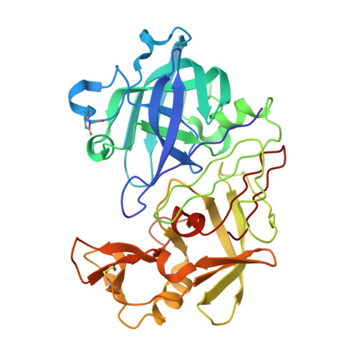
    The X-ray structures of native endothiapepsin and a complex with a hydroxyethylene transition state analog inhibitor (H261) have been determined at atomic resolution. Unrestrained refinement of the carboxyl groups of the enzyme by using the atomic resolution data indicates that both catalytic aspartates in the native enzyme share a single negative charge equally; that is, in the crystal, one half of the active sites have Asp 32 ionized and the other half have Asp 215 ionized. The electron density map of the native enzyme refined at 0.9 A resolution demonstrates that there is a short peptide (probably Ser-Thr) bound noncovalently in the active site cleft. The N-terminal nitrogen of the dipeptide interacts with the aspartate diad of the enzyme by hydrogen bonds involving the carboxyl of Asp 215 and the catalytic water molecule. This is consistent with classical findings that the aspartic proteinases can be inhibited weakly by short peptides and that these enzymes can catalyze transpeptidation reactions. The dipeptide may originate from autolysis of the N-terminal Ser-Thr sequence of the enzyme during crystallization.


  • Organizational Affiliation
    • Division of Biochemistry and Molecular Biology, School of Biological Sciences, University of Southampton, Bassett Crescent East, Southampton SO16 7PX, UK.

Macromolecule Content 

  • Total Structure Weight: 34.59 kDa 
  • Atom Count: 3,134 
  • Modeled Residue Count: 329 
  • Deposited Residue Count: 329 
  • Unique protein chains: 1

Macromolecules

Find similar proteins by:|  3D Structure
Entity ID: 1
MoleculeChains  Sequence LengthOrganismDetailsImage
ENDOTHIAPEPSIN329Cryphonectria parasiticaMutation(s): 0 
EC: 3.4.23.22
UniProt
Find proteins for P11838 (Cryphonectria parasitica)
Explore P11838 
Go to UniProtKB:  P11838
Entity Groups
Sequence Clusters30% Identity50% Identity70% Identity90% Identity95% Identity100% Identity
UniProt GroupP11838
Sequence Annotations
Expand
Reference Sequence

Small Molecules

Ligands 4 Unique
IDChains Name / Formula / InChI Key2D Diagram3D Interactions
THR

Query on THR


Download:Ideal Coordinates CCD File
C [auth A]THREONINE
C4 H9 N O3
AYFVYJQAPQTCCC-GBXIJSLDSA-N
SER

Query on SER


Download:Ideal Coordinates CCD File
B [auth A]SERINE
C3 H7 N O3
MTCFGRXMJLQNBG-REOHCLBHSA-N
SO4

Query on SO4


Download:Ideal Coordinates CCD File
E [auth A],
F [auth A],
G [auth A],
H [auth A],
I [auth A]
SULFATE ION
O4 S
QAOWNCQODCNURD-UHFFFAOYSA-L
GOL

Query on GOL


Download:Ideal Coordinates CCD File
D [auth A]GLYCEROL
C3 H8 O3
PEDCQBHIVMGVHV-UHFFFAOYSA-N
Modified Residues  1 Unique
IDChains TypeFormula2D DiagramParent
SUI
Query on SUI
A
L-PEPTIDE LINKINGC6 H8 N2 O4ASP, GLY

Experimental Data & Validation

Experimental Data

  • Method: X-RAY DIFFRACTION
  • Resolution: 0.90 Å
  • R-Value Free:  0.147 (Depositor) 
  • R-Value Observed: 0.121 (Depositor) 
Space Group: P 1 21 1
Unit Cell:
Length ( Å )Angle ( ˚ )
a = 42.72α = 90
b = 74.66β = 97.27
c = 42.55γ = 90
Software Package:
Software NamePurpose
SHELXL-97refinement
MOSFLMdata reduction
SCALAdata scaling
SHELXL-97phasing

Structure Validation

View Full Validation Report


Entry History 

Deposition Data

Revision History  (Full details and data files)

  • Version 1.0: 2003-04-02
    Type: Initial release
  • Version 1.1: 2011-05-08
    Changes: Version format compliance
  • Version 1.2: 2011-07-13
    Changes: Version format compliance
  • Version 1.3: 2018-06-20
    Changes: Advisory, Data collection, Derived calculations
  • Version 1.4: 2019-07-10
    Changes: Data collection
  • Version 1.5: 2019-07-24
    Changes: Data collection
  • Version 2.0: 2023-11-15
    Changes: Atomic model, Data collection, Database references, Derived calculations, Other