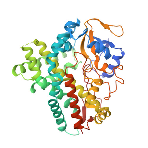
Comparison of the 1.85 A Structure of Cyp154A1 from Streptomyces Coelicolor A3(2) with the Closely Related Cyp154C1 and Cyps from Antibiotic Biosynthetic Pathways.
Podust, L.M., Bach, H., Kim, Y., Lamb, D.C., Arase, M., Sherman, D.H., Kelly, S.L., Waterman, M.R.(2004) Protein Sci 13: 255
- PubMed: 14691240
- DOI: https://doi.org/10.1110/ps.03384804
- Primary Citation of Related Structures:
1ODO - PubMed Abstract:
The genus Streptomyces produces two-thirds of microbially derived antibiotics. Polyketides form the largest and most diverse group of these natural products. Antibiotic diversity of polyketides is generated during their biosynthesis by several means, including postpolyketide modification performed by oxidoreductases, a broad group of enzymes including cytochrome P450 monooxygenases (CYPs). CYPs catalyze site-specific oxidation of macrolide antibiotic precursors significantly affecting antibiotic activity. Efficient manipulation of Streptomyces CYPs in generating new antibiotics will require identification and/or engineering of monooxygenases with activities toward a diverse array of chemical substrates. To begin to link structure to function of CYPs involved in secondary metabolic pathways of industrially important species, we determined the X-ray structure of Streptomyces coelicolor A3(2) CYP154A1 at 1.85 A and analyzed it in the context of the closely related CYP154C1 and more distant CYPs from polyketide synthase (EryF) and nonribosomal peptide synthetase (OxyB) biosynthetic pathways. In contrast to CYP154C1, CYP154A1 reveals an active site inaccessible from the molecular surface, and an absence of catalytic activities observed for CYP154C1. Systematic variations in the amino acid patterns and length of the surface HI loop correlate with degree of rotation of the F and G helices relative to the active site in CYP154A1-related CYPs, presumably regulating the degree of active site accessibility and its dimensions. Heme in CYP154A1 is in a 180 degrees flipped orientation compared with most other structurally determined CYPs.
Organizational Affiliation:
Department of Biochemistry, Vanderbilt University School of Medicine, Nashville, Tennessee 37232-0146, USA. larissa.m.podust@vanderbuilt.edu


