1OD5 | pdb_00001od5

Crystal structure of glycinin A3B4 subunit homohexamer


Experimental Data Snapshot

  • Method: X-RAY DIFFRACTION
  • Resolution: 2.10 Å
  • R-Value Free: 
    0.254 (Depositor), 0.243 (DCC) 
  • R-Value Work: 
    0.201 (Depositor), 0.193 (DCC) 
  • R-Value Observed: 
    0.201 (Depositor) 

Starting Model: experimental
View more details

wwPDB Validation 3D Report Full Report

Validation slider image for 1OD5

This is version 1.4 of the entry. See complete history

Literature

Crystal Structure of Soybean 11S Globulin: Glycinin A3B4 Homohexamer

Adachi, M.Kanamori, J.Masuda, T.Yagasaki, K.Kitamura, K.Mikami, B.Utsumi, S.

(2003) Proc Natl Acad Sci U S A 100: 7395

  • DOI: https://doi.org/10.1073/pnas.0832158100
  • Primary Citation Related Structures: 
    1OD5

  • PubMed Abstract: 

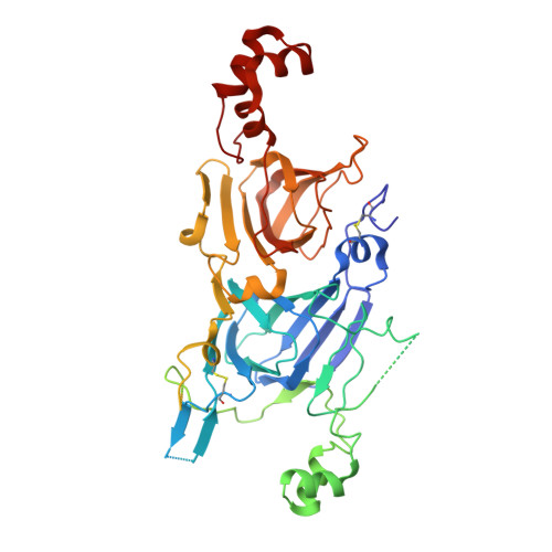
    Most plant seeds contain 11S globulins as major storage proteins for their nutrition. Soybean glycinin belongs to the 11S globulin family and consists of five kinds of subunits. We determined the crystal structure of a homohexamer of the glycinin A3B4 subunit at 2.1-A resolution. The crystal structure shows that the hexamer has 32-point group symmetry formed by face-to-face stacking of two trimers. The interface buries the highly conserved interchain disulfide. Based on the structure, we propose that an ingenious face-to-face mechanism controls the hexamer formation of the 11S globulin by movement of a mobile disordered region to the side of the trimer after posttranslational processing. Electrostatic analysis of the faces suggests that the interchain disulfide-containing face has high positive potential at acidic pH, which induces dissociation of the hexamer into trimers that may be susceptible to proteinases after seed imbibition. This dissociation might result in the degradation and mobilization of 11S globulins as storage proteins in embryos during germination and seedling growth.


  • Organizational Affiliation
    • Laboratory of Food Quality Design and Development, Graduate School of Agriculture, Kyoto University, Uji, Kyoto 611-0011, Japan.

Macromolecule Content 

  • Total Structure Weight: 111.38 kDa 
  • Atom Count: 6,220 
  • Modeled Residue Count: 764 
  • Deposited Residue Count: 984 
  • Unique protein chains: 1

Macromolecules

Find similar proteins by:|  3D Structure
Entity ID: 1
MoleculeChains  Sequence LengthOrganismDetailsImage
GLYCININ
A, B
492Glycine maxMutation(s): 0 
UniProt
Find proteins for P04347 (Glycine max)
Explore P04347 
Go to UniProtKB:  P04347
Entity Groups
Sequence Clusters30% Identity50% Identity70% Identity90% Identity95% Identity100% Identity
UniProt GroupP04347
Sequence Annotations
Expand
Reference Sequence

Experimental Data & Validation

Experimental Data

  • Method: X-RAY DIFFRACTION
  • Resolution: 2.10 Å
  • R-Value Free:  0.254 (Depositor), 0.243 (DCC) 
  • R-Value Work:  0.201 (Depositor), 0.193 (DCC) 
  • R-Value Observed: 0.201 (Depositor) 
Space Group: H 3
Unit Cell:
Length ( Å )Angle ( ˚ )
a = 114.842α = 90
b = 114.842β = 90
c = 191.572γ = 120
Software Package:
Software NamePurpose
X-PLORrefinement
SAINTdata reduction
SAINTdata scaling
X-PLORphasing

Structure Validation

View Full Validation Report


Entry History 

Deposition Data

Revision History  (Full details and data files)

  • Version 1.0: 2003-06-02
    Type: Initial release
  • Version 1.1: 2015-06-10
    Changes: Database references, Derived calculations, Non-polymer description, Other, Version format compliance
  • Version 1.2: 2018-04-04
    Changes: Data collection
  • Version 1.3: 2023-12-13
    Changes: Data collection, Database references, Derived calculations, Other, Refinement description
  • Version 1.4: 2024-10-23
    Changes: Structure summary