1OBP | pdb_00001obp

ODORANT-BINDING PROTEIN FROM BOVINE NASAL MUCOSA


Experimental Data Snapshot

  • Method: X-RAY DIFFRACTION
  • Resolution: 2.00 Å
  • R-Value Free: 
    0.247 (Depositor) 
  • R-Value Work: 
    0.164 (Depositor), 0.223 (DCC) 
  • R-Value Observed: 
    0.164 (Depositor) 

wwPDB Validation 3D Report Full Report

Validation slider image for 1OBP

This is version 1.3 of the entry. See complete history

Literature

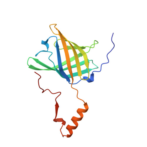
Domain swapping creates a third putative combining site in bovine odorant binding protein dimer

Tegoni, M.Ramoni, R.Bignetti, E.Spinelli, S.Cambillau, C.

(1996) Nat Struct Biol 3: 863-867

  • DOI: https://doi.org/10.1038/nsb1096-863
  • Primary Citation Related Structures: 
    1OBP

  • PubMed Abstract: 

    In mammals, odorant binding proteins may play an important role in the transport of odors towards specific olfactory receptors on sensory neurones across the aqueous compartment of the nasal mucus. We have solved the X-ray structure of such a transport protein, bovine odorant binding protein (OBP) at 2.0 A resolution. The beta-barrel of OBP is similar to that of lipocalins, but OBP dimer association results from domain swapping, an observation unique among the lipocalins. The alpha-helix of each monomer stacks against the beta-barrel of the other monomer. Contrary to previous reports, each monomer has an internal buried cavity which could accommodate a naturally occurring molecule. Besides this cavity, an open cavity is located at the dimer interface. Data in solution suggest that this central cavity may be a binding site created by domain swapping.


  • Organizational Affiliation
    • UPR9039-CNRS, IFR1, Marseille, France.

Macromolecule Content 

  • Total Structure Weight: 36.91 kDa 
  • Atom Count: 2,914 
  • Modeled Residue Count: 313 
  • Deposited Residue Count: 318 
  • Unique protein chains: 1

Macromolecules

Find similar proteins by:|  3D Structure
Entity ID: 1
MoleculeChains  Sequence LengthOrganismDetailsImage
ODORANT-BINDING PROTEIN
A, B
159Bos taurusMutation(s): 0 
UniProt
Find proteins for P07435 (Bos taurus)
Explore P07435 
Go to UniProtKB:  P07435
Entity Groups
Sequence Clusters30% Identity50% Identity70% Identity90% Identity95% Identity100% Identity
UniProt GroupP07435
Sequence Annotations
Expand
Reference Sequence

Small Molecules

Ligands 1 Unique
IDChains Name / Formula / InChI Key2D Diagram3D Interactions
UNX

Query on UNX


Download:Ideal Coordinates CCD File
AA [auth A]
AB [auth B]
AC [auth B]
BA [auth A]
BB [auth B]
AA [auth A],
AB [auth B],
AC [auth B],
BA [auth A],
BB [auth B],
BC [auth B],
C [auth A],
CA [auth A],
CB [auth B],
CC [auth B],
D [auth A],
DA [auth A],
DB [auth B],
DC [auth B],
E [auth A],
EA [auth A],
EB [auth B],
EC [auth B],
F [auth A],
FA [auth A],
FB [auth B],
FC [auth B],
G [auth A],
GA [auth A],
GB [auth B],
H [auth A],
HA [auth A],
HB [auth B],
I [auth A],
IA [auth A],
IB [auth B],
J [auth A],
JA [auth A],
JB [auth B],
K [auth A],
KA [auth A],
KB [auth B],
L [auth A],
LA [auth A],
LB [auth B],
M [auth A],
MA [auth A],
MB [auth B],
N [auth A],
NA [auth A],
NB [auth B],
O [auth A],
OA [auth A],
OB [auth B],
P [auth A],
PA [auth A],
PB [auth B],
Q [auth A],
QA [auth A],
QB [auth B],
R [auth A],
RA [auth B],
RB [auth B],
S [auth A],
SA [auth B],
SB [auth B],
T [auth A],
TA [auth B],
TB [auth B],
U [auth A],
UA [auth B],
UB [auth B],
V [auth A],
VA [auth B],
VB [auth B],
W [auth A],
WA [auth B],
WB [auth B],
X [auth A],
XA [auth B],
XB [auth B],
Y [auth A],
YA [auth B],
YB [auth B],
Z [auth A],
ZA [auth B],
ZB [auth B]
UNKNOWN ATOM OR ION
X

Experimental Data & Validation

Experimental Data

  • Method: X-RAY DIFFRACTION
  • Resolution: 2.00 Å
  • R-Value Free:  0.247 (Depositor) 
  • R-Value Work:  0.164 (Depositor), 0.223 (DCC) 
  • R-Value Observed: 0.164 (Depositor) 
Space Group: P 1 21 1
Unit Cell:
Length ( Å )Angle ( ˚ )
a = 55.9α = 90
b = 65.5β = 98.8
c = 42.7γ = 90
Software Package:
Software NamePurpose
X-PLORmodel building
X-PLORrefinement
DENZOdata reduction
X-PLORphasing

Structure Validation

View Full Validation Report


Entry History 

Deposition Data

Revision History  (Full details and data files)

  • Version 1.0: 1996-10-14
    Type: Initial release
  • Version 1.1: 2008-03-24
    Changes: Version format compliance
  • Version 1.2: 2011-07-13
    Changes: Version format compliance
  • Version 1.3: 2024-02-14
    Changes: Data collection, Database references, Derived calculations, Other