1OAS | pdb_00001oas

O-ACETYLSERINE SULFHYDRYLASE FROM SALMONELLA TYPHIMURIUM


Experimental Data Snapshot

  • Method: X-RAY DIFFRACTION
  • Resolution: 2.20 Å
  • R-Value Free: 
    0.200 (Depositor), 0.201 (DCC) 
  • R-Value Work: 
    0.172 (Depositor), 0.173 (DCC) 

wwPDB Validation 3D Report Full Report

Validation slider image for 1OAS

Ligand Structure Quality Assessment 


This is version 1.3 of the entry. See complete history

Literature

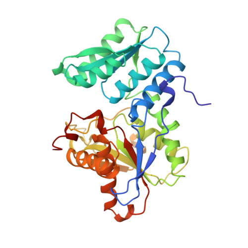
Three-dimensional structure of O-acetylserine sulfhydrylase from Salmonella typhimurium.

Burkhard, P.Rao, G.S.Hohenester, E.Schnackerz, K.D.Cook, P.F.Jansonius, J.N.

(1998) J Mol Biology 283: 121-133

  • DOI: https://doi.org/10.1006/jmbi.1998.2037
  • Primary Citation Related Structures: 
    1OAS

  • PubMed Abstract: 

    The last step in cysteine biosynthesis in enteric bacteria is catalyzed by the pyridoxal 5'-phosphate-dependent enzyme O-acetylserine sulfhydrylase. Here we report the crystal structure at 2.2 A resolution of the A-isozyme of O-acetylserine sulfhydrylase isolated from Salmonella typhimurium. O-acetylserine sulfhydrylase shares the same fold with tryptophan synthase-beta from Salmonella typhimurium but the sequence identity level is below 20%. There are some major structural differences: the loops providing the interface to the alpha-subunit in tryptophan synthase-beta and two surface helices of tryptophan synthase-beta are missing in O-acetylserine sulfhydrylase. The hydrophobic channel for indole transport from the alpha to the beta active site of tryptophan synthase-beta is, not unexpectedly, also absent in O-acetylserine sulfhydrylase. The dimer interface, on the other hand, is more or less conserved in the two enzymes. The active site cleft of O-acetylserine sulfhydrylase is wider and therefore more exposed to the solvent. A possible binding site for the substrate O-acetylserine is discussed.


  • Organizational Affiliation
    • Department of Structural Biology, Biozentrum, University of Basel, Klingelbergstrasse70, Basel, CH-4056, Switzerland.

Macromolecule Content 

  • Total Structure Weight: 69.44 kDa 
  • Atom Count: 5,158 
  • Modeled Residue Count: 630 
  • Deposited Residue Count: 644 
  • Unique protein chains: 1

Macromolecules

Find similar proteins by:|  3D Structure
Entity ID: 1
MoleculeChains  Sequence LengthOrganismDetailsImage
O-ACETYLSERINE SULFHYDRYLASE
A, B
322Salmonella enterica subsp. enterica serovar TyphimuriumMutation(s): 0 
EC: 4.2.99.8 (PDB Primary Data), 2.5.1.47 (UniProt)
UniProt
Find proteins for P0A1E3 (Salmonella typhimurium (strain LT2 / SGSC1412 / ATCC 700720))
Explore P0A1E3 
Go to UniProtKB:  P0A1E3
Entity Groups
Sequence Clusters30% Identity50% Identity70% Identity90% Identity95% Identity100% Identity
UniProt GroupP0A1E3
Sequence Annotations
Expand
Reference Sequence

Experimental Data & Validation

Experimental Data

  • Method: X-RAY DIFFRACTION
  • Resolution: 2.20 Å
  • R-Value Free:  0.200 (Depositor), 0.201 (DCC) 
  • R-Value Work:  0.172 (Depositor), 0.173 (DCC) 
Space Group: P 21 21 21
Unit Cell:
Length ( Å )Angle ( ˚ )
a = 54.6α = 90
b = 98.2β = 90
c = 145.4γ = 90
Software Package:
Software NamePurpose
MLPHAREphasing
CNSrefinement
MOSFLMdata reduction
CCP4data scaling

Structure Validation

View Full Validation Report


Ligand Structure Quality Assessment 


Entry History 

Deposition Data

Revision History  (Full details and data files)

  • Version 1.0: 2000-01-28
    Type: Initial release
  • Version 1.1: 2007-10-16
    Changes: Version format compliance
  • Version 1.2: 2011-07-13
    Changes: Derived calculations, Version format compliance
  • Version 1.3: 2023-12-27
    Changes: Data collection, Database references, Derived calculations