1OAR | pdb_00001oar

Fv IgE SPE-7 in complex with Alizarin Red


Experimental Data Snapshot

  • Method: X-RAY DIFFRACTION
  • Resolution: 2.23 Å
  • R-Value Free: 
    0.241 (Depositor), 0.231 (DCC) 
  • R-Value Work: 
    0.205 (Depositor), 0.235 (DCC) 

Starting Model: experimental
View more details

wwPDB Validation 3D Report Full Report

Validation slider image for 1OAR

Ligand Structure Quality Assessment 


This is version 1.3 of the entry. See complete history

Literature

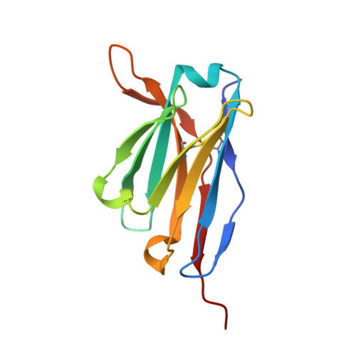
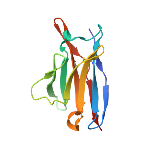
Antibody Multispecificity Mediated by Conformational Diversity

James, L.C.Roversi, P.Tawfik, D.

(2003) Science 299: 1362

  • DOI: https://doi.org/10.1126/science.1079731
  • Primary Citation Related Structures: 
    1OAQ, 1OAR, 1OAU, 1OAX, 1OAY, 1OAZ, 1OCW

  • PubMed Abstract: 

    A single antibody was shown to adopt different binding-site conformations and thereby bind unrelated antigens. Analysis by both x-ray crystallography and pre-steady-state kinetics revealed an equilibrium between different preexisting isomers, one of which possessed a promiscuous, low-affinity binding site for aromatic ligands, including the immunizing hapten. A subsequent induced-fit isomerization led to high-affinity complexes with a deep and narrow binding site. A protein antigen identified by repertoire selection made use of an unrelated antibody isomer with a wide, shallow binding site. Conformational diversity, whereby one sequence adopts multiple structures and multiple functions, can increase the effective size of the antibody repertoire but may also lead to autoimmunity and allergy.


  • Organizational Affiliation
    • Centre for Protein Engineering, Medical Research Council Centre, Hills Road, Cambridge CB2 2HQ, UK.

Macromolecule Content 

  • Total Structure Weight: 104.85 kDa 
  • Atom Count: 7,050 
  • Modeled Residue Count: 839 
  • Deposited Residue Count: 928 
  • Unique protein chains: 2

Macromolecules

Find similar proteins by:|  3D Structure
Entity ID: 1
MoleculeChains  Sequence LengthOrganismDetailsImage
IMMUNOGLOBULIN EA [auth H],
B [auth I],
C [auth J],
D [auth K]
122Mus musculusMutation(s): 0 
Find similar proteins by:|  3D Structure
Entity ID: 2
MoleculeChains  Sequence LengthOrganismDetailsImage
IMMUNOGLOBULING EE [auth L],
F [auth M],
G [auth N],
H [auth O]
110Mus musculusMutation(s): 0 
UniProt
Find proteins for P01724 (Mus musculus)
Explore P01724 
Go to UniProtKB:  P01724
Entity Groups
Sequence Clusters30% Identity50% Identity70% Identity90% Identity95% Identity100% Identity
UniProt GroupP01724
Sequence Annotations
Expand
Reference Sequence

Small Molecules

Ligands 6 Unique
IDChains Name / Formula / InChI Key2D Diagram3D Interactions
AZN

Query on AZN


Download:Ideal Coordinates CCD File
EA [auth J],
HA [auth K],
R [auth H],
T [auth I]
ALIZARIN RED
C14 H8 O7 S
JKYKXTRKURYNGW-UHFFFAOYSA-N
CAC

Query on CAC


Download:Ideal Coordinates CCD File
I [auth H],
V [auth J]
CACODYLATE ION
C2 H6 As O2
OGGXGZAMXPVRFZ-UHFFFAOYSA-M
DMS

Query on DMS


Download:Ideal Coordinates CCD File
AA [auth J]
BA [auth J]
CA [auth J]
IA [auth L]
J [auth H]
AA [auth J],
BA [auth J],
CA [auth J],
IA [auth L],
J [auth H],
JA [auth L],
K [auth H],
KA [auth L],
L [auth H],
LA [auth L],
M [auth H],
MA [auth L],
N [auth H],
O [auth H],
P [auth H],
TA [auth N],
UA [auth N],
VA [auth N],
W [auth J],
WA [auth N],
X [auth J],
XA [auth N],
Y [auth J],
Z [auth J]
DIMETHYL SULFOXIDE
C2 H6 O S
IAZDPXIOMUYVGZ-UHFFFAOYSA-N
EDO

Query on EDO


Download:Ideal Coordinates CCD File
DA [auth J],
Q [auth H]
1,2-ETHANEDIOL
C2 H6 O2
LYCAIKOWRPUZTN-UHFFFAOYSA-N
CL

Query on CL


Download:Ideal Coordinates CCD File
AB [auth N]
BB [auth O]
GA [auth J]
OA [auth L]
PA [auth L]
AB [auth N],
BB [auth O],
GA [auth J],
OA [auth L],
PA [auth L],
QA [auth L],
SA [auth M],
ZA [auth N]
CHLORIDE ION
Cl
VEXZGXHMUGYJMC-UHFFFAOYSA-M
NA

Query on NA


Download:Ideal Coordinates CCD File
FA [auth J]
NA [auth L]
RA [auth M]
S [auth H]
U [auth I]
FA [auth J],
NA [auth L],
RA [auth M],
S [auth H],
U [auth I],
YA [auth N]
SODIUM ION
Na
FKNQFGJONOIPTF-UHFFFAOYSA-N

Experimental Data & Validation

Experimental Data

  • Method: X-RAY DIFFRACTION
  • Resolution: 2.23 Å
  • R-Value Free:  0.241 (Depositor), 0.231 (DCC) 
  • R-Value Work:  0.205 (Depositor), 0.235 (DCC) 
Space Group: P 21 21 21
Unit Cell:
Length ( Å )Angle ( ˚ )
a = 78.917α = 90
b = 78.882β = 90
c = 169.036γ = 90
Software Package:
Software NamePurpose
REFMACrefinement
MOSFLMdata reduction
SCALAdata scaling
AMoREphasing

Structure Validation

View Full Validation Report


Ligand Structure Quality Assessment 


Entry History 

Deposition Data

Revision History  (Full details and data files)

  • Version 1.0: 2004-01-15
    Type: Initial release
  • Version 1.1: 2013-10-30
    Changes: Derived calculations, Other, Source and taxonomy, Version format compliance
  • Version 1.2: 2024-11-13
    Changes: Data collection, Database references, Derived calculations, Other, Structure summary
  • Version 1.3: 2026-03-04
    Changes: Refinement description, Structure summary