1OA8 | pdb_00001oa8

AXH domain of human spinocerebellar ataxin-1


Experimental Data Snapshot

  • Method: X-RAY DIFFRACTION
  • Resolution: 1.70 Å
  • R-Value Free: 
    0.252 (Depositor) 
  • R-Value Work: 
    0.210 (Depositor) 

wwPDB Validation 3D Report Full Report

Validation slider image for 1OA8

This is version 1.6 of the entry. See complete history

Literature

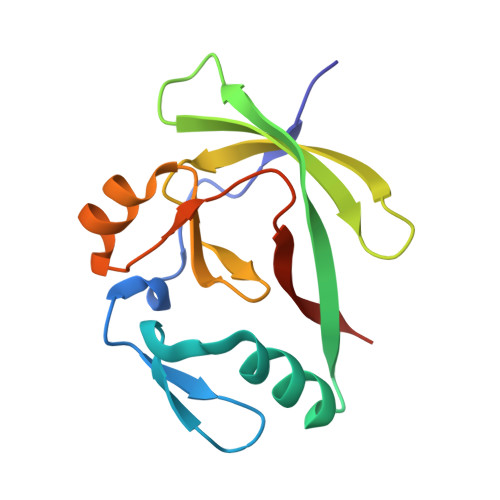
The structure of the AXH domain of spinocerebellar ataxin-1.

Chen, Y.W.Allen, M.D.Veprintsev, D.B.Lowe, J.Bycroft, M.

(2004) J Biological Chem 279: 3758-3765

  • DOI: https://doi.org/10.1074/jbc.M309817200
  • Primary Citation Related Structures: 
    1OA8

  • PubMed Abstract: 

    Spinocerebellar ataxia type 1 is a late-onset neurodegenerative disease caused by the expansion of a CAG triplet repeat in the SCA1 gene. This results in the lengthening of a polyglutamine tract in the gene product ataxin-1. This produces a toxic gain of function that results in specific neuronal death. A region in ataxin-1, the AXH domain, exhibits significant sequence similarity to the transcription factor HBP1. This region of the protein has been implicated in RNA binding and self-association. We have determined the crystal structure of the AXH domain of ataxin-1. The AXH domain is dimeric and contains an OB-fold, a structural motif found in many oligonucleotide-binding proteins, supporting its proposed role in RNA binding. By structure comparison with other proteins that contain an OB-fold, a putative RNA-binding site has been identified. We also identified a cluster of charged surface residues that are well conserved among AXH domains. These residues may constitute a second ligand-binding surface, suggesting that all AXH domains interact with a common yet unidentified partner.


  • Organizational Affiliation
    • Centre for Protein Engineering, Medical Research Council Centre, Cambridge, UK.

Macromolecule Content 

  • Total Structure Weight: 57.78 kDa 
  • Atom Count: 4,315 
  • Modeled Residue Count: 518 
  • Deposited Residue Count: 532 
  • Unique protein chains: 1

Macromolecules

Find similar proteins by:|  3D Structure
Entity ID: 1
MoleculeChains  Sequence LengthOrganismDetailsImage
ATAXIN-1
A, B, C, D
133Homo sapiensMutation(s): 0 
UniProt & NIH Common Fund Data Resources
Find proteins for P54253 (Homo sapiens)
Explore P54253 
Go to UniProtKB:  P54253
PHAROS:  P54253
GTEx:  ENSG00000124788 
Entity Groups
Sequence Clusters30% Identity50% Identity70% Identity90% Identity95% Identity100% Identity
UniProt GroupP54253
Sequence Annotations
Expand
Reference Sequence

Experimental Data & Validation

Experimental Data

  • Method: X-RAY DIFFRACTION
  • Resolution: 1.70 Å
  • R-Value Free:  0.252 (Depositor) 
  • R-Value Work:  0.210 (Depositor) 
Space Group: P 21 21 21
Unit Cell:
Length ( Å )Angle ( ˚ )
a = 48.783α = 90
b = 82.441β = 90
c = 137.781γ = 90
Software Package:
Software NamePurpose
REFMACrefinement
MOSFLMdata reduction
SCALAdata scaling
SOLVEphasing

Structure Validation

View Full Validation Report


Entry History 

Deposition Data

Revision History  (Full details and data files)

  • Version 1.0: 2003-11-06
    Type: Initial release
  • Version 1.1: 2011-05-08
    Changes: Version format compliance
  • Version 1.2: 2011-07-13
    Changes: Version format compliance
  • Version 1.3: 2018-01-24
    Changes: Source and taxonomy
  • Version 1.4: 2019-02-06
    Changes: Data collection, Database references, Experimental preparation
  • Version 1.5: 2019-10-09
    Changes: Data collection, Derived calculations
  • Version 1.6: 2024-05-08
    Changes: Data collection, Database references, Derived calculations