1O9W | pdb_00001o9w

F17-aG lectin domain from Escherichia coli in complex with N-acetyl-glucosamine


Experimental Data Snapshot

  • Method: X-RAY DIFFRACTION
  • Resolution: 1.65 Å
  • R-Value Free: 
    0.242 (Depositor), 0.398 (DCC) 
  • R-Value Work: 
    0.211 (Depositor), 0.403 (DCC) 
  • R-Value Observed: 
    0.211 (Depositor) 

Starting Model: experimental
View more details

wwPDB Validation 3D Report Full Report

Validation slider image for 1O9W

Ligand Structure Quality Assessment 


This is version 1.5 of the entry. See complete history

Literature

The Fimbrial Adhesin F17-G of Enterotoxigenic Escherichia Coli Has an Immunoglobulin-Like Lectin Domain that Binds N-Acetylglucosamine

Buts, L.Bouckaert, J.De Genst, E.Loris, R.Oscarson, S.Lahmann, M.Messens, J.Brosens, E.Wyns, L.De Greve, H.

(2003) Mol Microbiol 49: 705

  • DOI: https://doi.org/10.1046/j.1365-2958.2003.03600.x
  • Primary Citation Related Structures: 
    1O9V, 1O9W, 1O9Z

  • PubMed Abstract: 

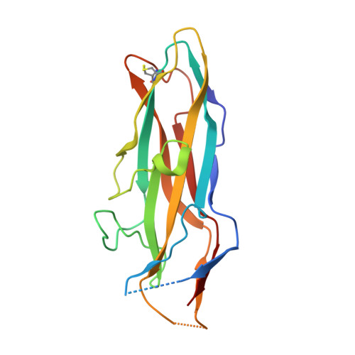
    The F17-G adhesin at the tip of flexible F17 fimbriae of enterotoxigenic Escherichia coli mediates binding to N-acetyl-beta-D-glucosamine-presenting receptors on the microvilli of the intestinal epithelium of ruminants. We report the 1.7 A resolution crystal structure of the lectin domain of F17-G, both free and in complex with N-acetylglucosamine. The monosaccharide is bound on the side of the ellipsoid-shaped protein in a conserved site around which all natural variations of F17-G are clustered. A model is proposed for the interaction between F17-fimbriated E. coli and microvilli with enhanced affinity compared with the binding constant we determined for F17-G binding to N-acetylglucosamine (0.85 mM-1). Unexpectedly, the F17-G structure reveals that the lectin domains of the F17-G, PapGII and FimH fimbrial adhesins all share the immunoglobulin-like fold of the structural components (pilins) of their fimbriae, despite lack of any sequence identity. Fold comparisons with pilin and chaperone structures of the chaperone/usher pathway highlight the central role of the C-terminal beta-strand G of the immunoglobulin-like fold and provides new insights into pilus assembly, function and adhesion.


  • Organizational Affiliation
    • Department of Ultrastructure, Institute for Molecular Biology, Vrije Universiteit Brussel, Vlaams Interuniversitair Instituut voor Biotechnologie, Brussels, Belgium.

Macromolecule Content 

  • Total Structure Weight: 19.27 kDa 
  • Atom Count: 1,370 
  • Modeled Residue Count: 169 
  • Deposited Residue Count: 177 
  • Unique protein chains: 1

Macromolecules

Find similar proteins by:|  3D Structure
Entity ID: 1
MoleculeChains  Sequence LengthOrganismDetailsImage
F17A-G FIMBRIAL ADHESIN177Escherichia coliMutation(s): 0 
UniProt
Find proteins for Q99003 (Escherichia coli)
Explore Q99003 
Go to UniProtKB:  Q99003
Entity Groups
Sequence Clusters30% Identity50% Identity70% Identity90% Identity95% Identity100% Identity
UniProt GroupQ99003
Sequence Annotations
Expand
Reference Sequence

Small Molecules

Ligands 1 Unique
IDChains Name / Formula / InChI Key2D Diagram3D Interactions
NAG

Query on NAG


Download:Ideal Coordinates CCD File
B [auth A]2-acetamido-2-deoxy-beta-D-glucopyranose
C8 H15 N O6
OVRNDRQMDRJTHS-FMDGEEDCSA-N

Experimental Data & Validation

Experimental Data

  • Method: X-RAY DIFFRACTION
  • Resolution: 1.65 Å
  • R-Value Free:  0.242 (Depositor), 0.398 (DCC) 
  • R-Value Work:  0.211 (Depositor), 0.403 (DCC) 
  • R-Value Observed: 0.211 (Depositor) 
Space Group: P 61 2 2
Unit Cell:
Length ( Å )Angle ( ˚ )
a = 42.329α = 90
b = 42.329β = 90
c = 268.714γ = 120
Software Package:
Software NamePurpose
CNSrefinement
DENZOdata reduction
SCALEPACKdata scaling
CNSphasing

Structure Validation

View Full Validation Report


Ligand Structure Quality Assessment 


Entry History 

Deposition Data

Revision History  (Full details and data files)

  • Version 1.0: 2003-05-29
    Type: Initial release
  • Version 1.1: 2012-01-18
    Changes: Database references, Derived calculations, Non-polymer description, Other, Refinement description, Structure summary, Version format compliance
  • Version 1.2: 2019-07-24
    Changes: Advisory, Data collection
  • Version 1.3: 2020-07-29
    Type: Remediation
    Reason: Carbohydrate remediation
    Changes: Data collection, Derived calculations, Other, Structure summary
  • Version 1.4: 2023-12-13
    Changes: Advisory, Data collection, Database references, Refinement description, Structure summary
  • Version 1.5: 2024-10-23
    Changes: Structure summary