1O8A | pdb_00001o8a

Crystal Structure of Human Angiotensin Converting Enzyme (Native).


Experimental Data Snapshot

  • Method: X-RAY DIFFRACTION
  • Resolution: 2.00 Å
  • R-Value Free: 
    0.220 (Depositor) 
  • R-Value Work: 
    0.180 (Depositor) 
  • R-Value Observed: 
    0.180 (Depositor) 

Starting Model: experimental
View more details

wwPDB Validation 3D Report Full Report

Validation slider image for 1O8A

This is version 2.3 of the entry. See complete history

Literature

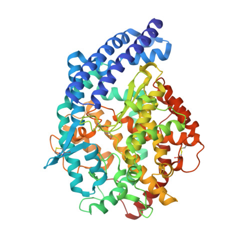
Crystal structure of the human angiotensin-converting enzyme-lisinopril complex.

Natesh, R.Schwager, S.L.Sturrock, E.D.Acharya, K.R.

(2003) Nature 421: 551-554

  • DOI: https://doi.org/10.1038/nature01370
  • Primary Citation Related Structures: 
    1O86, 1O8A

  • PubMed Abstract: 

    Angiotensin-converting enzyme (ACE) has a critical role in cardiovascular function by cleaving the carboxy terminal His-Leu dipeptide from angiotensin I to produce a potent vasopressor octapeptide, angiotensin II. Inhibitors of ACE are a first line of therapy for hypertension, heart failure, myocardial infarction and diabetic nephropathy. Notably, these inhibitors were developed without knowledge of the structure of human ACE, but were instead designed on the basis of an assumed mechanistic homology with carboxypeptidase A. Here we present the X-ray structure of human testicular ACE and its complex with one of the most widely used inhibitors, lisinopril (N2-[(S)-1-carboxy-3-phenylpropyl]-L-lysyl-L-proline; also known as Prinivil or Zestril), at 2.0 A resolution. Analysis of the three-dimensional structure of ACE shows that it bears little similarity to that of carboxypeptidase A, but instead resembles neurolysin and Pyrococcus furiosus carboxypeptidase--zinc metallopeptidases with no detectable sequence similarity to ACE. The structure provides an opportunity to design domain-selective ACE inhibitors that may exhibit new pharmacological profiles.


  • Organizational Affiliation
    • Department of Biology and Biochemistry, University of Bath, Claverton Down, Bath BA2 7AY, UK.

Macromolecule Content 

  • Total Structure Weight: 69.72 kDa 
  • Atom Count: 5,264 
  • Modeled Residue Count: 575 
  • Deposited Residue Count: 589 
  • Unique protein chains: 1

Macromolecules

Find similar proteins by:|  3D Structure
Entity ID: 1
MoleculeChains  Sequence LengthOrganismDetailsImage
ANGIOTENSIN CONVERTING ENZYME589Homo sapiensMutation(s): 0 
EC: 3.4.15.1
UniProt & NIH Common Fund Data Resources
Find proteins for P12821 (Homo sapiens)
Explore P12821 
Go to UniProtKB:  P12821
PHAROS:  P12821
GTEx:  ENSG00000159640 
Entity Groups
Sequence Clusters30% Identity50% Identity70% Identity90% Identity95% Identity100% Identity
UniProt GroupP12821
Glycosylation
Glycosylation Sites: 6Go to GlyGen: P12821-1
Sequence Annotations
Expand
Reference Sequence

Small Molecules

Ligands 5 Unique
IDChains Name / Formula / InChI Key2D Diagram3D Interactions
NAG

Query on NAG


Download:Ideal Coordinates CCD File
B [auth A]
C [auth A]
D [auth A]
E [auth A]
F [auth A]
B [auth A],
C [auth A],
D [auth A],
E [auth A],
F [auth A],
G [auth A]
2-acetamido-2-deoxy-beta-D-glucopyranose
C8 H15 N O6
OVRNDRQMDRJTHS-FMDGEEDCSA-N
NXA

Query on NXA


Download:Ideal Coordinates CCD File
J [auth A]N-CARBOXYALANINE
C4 H7 N O4
JCVPWRQVQNVNGU-REOHCLBHSA-N
ZN

Query on ZN


Download:Ideal Coordinates CCD File
I [auth A]ZINC ION
Zn
PTFCDOFLOPIGGS-UHFFFAOYSA-N
ACT

Query on ACT


Download:Ideal Coordinates CCD File
H [auth A]ACETATE ION
C2 H3 O2
QTBSBXVTEAMEQO-UHFFFAOYSA-M
CL

Query on CL


Download:Ideal Coordinates CCD File
K [auth A],
L [auth A]
CHLORIDE ION
Cl
VEXZGXHMUGYJMC-UHFFFAOYSA-M

Experimental Data & Validation

Experimental Data

  • Method: X-RAY DIFFRACTION
  • Resolution: 2.00 Å
  • R-Value Free:  0.220 (Depositor) 
  • R-Value Work:  0.180 (Depositor) 
  • R-Value Observed: 0.180 (Depositor) 
Space Group: P 21 21 21
Unit Cell:
Length ( Å )Angle ( ˚ )
a = 56.62α = 90
b = 85.06β = 90
c = 133.79γ = 90
Software Package:
Software NamePurpose
CNSrefinement
HKL-2000data reduction
SCALEPACKdata scaling
CNSphasing

Structure Validation

View Full Validation Report


Entry History 

Deposition Data

Revision History  (Full details and data files)

  • Version 1.0: 2003-02-07
    Type: Initial release
  • Version 1.1: 2011-05-08
    Changes: Version format compliance
  • Version 1.2: 2011-07-13
    Changes: Version format compliance
  • Version 2.0: 2018-07-11
    Changes: Advisory, Atomic model, Data collection, Database references, Derived calculations, Non-polymer description, Structure summary
  • Version 2.1: 2020-07-29
    Type: Remediation
    Reason: Carbohydrate remediation
    Changes: Data collection, Derived calculations, Structure summary
  • Version 2.2: 2023-12-13
    Changes: Data collection, Database references, Refinement description, Structure summary
  • Version 2.3: 2025-10-01
    Changes: Advisory, Derived calculations, Structure summary