1O7K

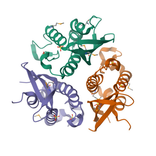
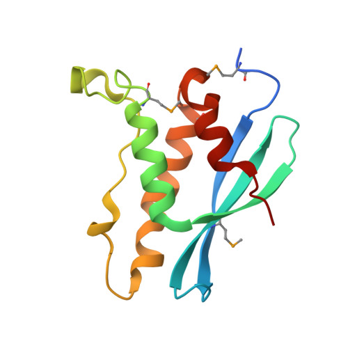
human p47 PX domain complex with sulphates


Experimental Data Snapshot

  • Method: X-RAY DIFFRACTION
  • Resolution: 2.00 Å
  • R-Value Free: 0.254 
  • R-Value Work: 0.210 
  • R-Value Observed: 0.223 

wwPDB Validation   3D Report Full Report


This is version 1.4 of the entry. See complete history


Literature

Binding of the Px Domain of P47Phox to Phosphatidylinositol 3.4-Bisphosphate and Phosphatidic Acid is Masked by an Intramolecular Interaction

Karathanassis, D.Stahelin, R.V.Bravo, J.Perisic, O.Pacold, C.M.Cho, W.Williams, R.L.

(2002) EMBO J 21: 5057

  • DOI: https://doi.org/10.1093/emboj/cdf519
  • Primary Citation of Related Structures:  
    1O7K

  • PubMed Abstract: 

    p47(phox) is a key cytosolic subunit required for activation of phagocyte NADPH oxidase. The X-ray structure of the p47(phox) PX domain revealed two distinct basic pockets on the membrane-binding surface, each occupied by a sulfate. These two pockets have different specificities: one preferentially binds phosphatidylinositol 3,4-bisphosphate [PtdIns(3,4)P(2)] and is analogous to the phophatidylinositol 3-phosphate (PtdIns3P)-binding pocket of p40(phox), while the other binds anionic phospholipids such as phosphatidic acid (PtdOH) or phosphatidylserine. The preference of this second site for PtdOH may be related to previously observed activation of NADPH oxidase by PtdOH. Simultaneous occupancy of the two phospholipid-binding pockets radically increases membrane affinity. Strikingly, measurements for full-length p47(phox) show that membrane interaction by the PX domain is masked by an intramolecular association with the C-terminal SH3 domain (C-SH3). Either a site-specific mutation in C-SH3 (W263R) or a mimic of the phosphorylated form of p47(phox) [Ser(303, 304, 328, 359, 370)Glu] cause a transition from a closed to an open conformation that binds membranes with a greater affinity than the isolated PX domain.


  • Organizational Affiliation

    MRC Laboratory of Molecular Biology, Hills Road, Cambridge CB2 2QH, UK.


Macromolecules
Find similar proteins by:  (by identity cutoff)  |  3D Structure
Entity ID: 1
MoleculeChains Sequence LengthOrganismDetailsImage
NEUTROPHIL CYTOSOL FACTOR 1
A, B, C
131Homo sapiensMutation(s): 0 
Membrane Entity: Yes 
UniProt & NIH Common Fund Data Resources
Find proteins for P14598 (Homo sapiens)
Explore P14598 
Go to UniProtKB:  P14598
PHAROS:  P14598
GTEx:  ENSG00000158517 
Entity Groups  
Sequence Clusters30% Identity50% Identity70% Identity90% Identity95% Identity100% Identity
UniProt GroupP14598
Sequence Annotations
Expand
  • Reference Sequence
Experimental Data & Validation

Experimental Data

  • Method: X-RAY DIFFRACTION
  • Resolution: 2.00 Å
  • R-Value Free: 0.254 
  • R-Value Work: 0.210 
  • R-Value Observed: 0.223 
  • Space Group: I 2 2 2
Unit Cell:
Length ( Å )Angle ( ˚ )
a = 74.233α = 90
b = 92.175β = 90
c = 144.01γ = 90
Software Package:
Software NamePurpose
REFMACrefinement
MOSFLMdata reduction
SCALAdata scaling
autoSHARPphasing
SHARPphasing
CCP4phasing

Structure Validation

View Full Validation Report


Entry History 

Deposition Data

Revision History  (Full details and data files)

  • Version 1.0: 2002-11-20
    Type: Initial release
  • Version 1.1: 2011-05-08
    Changes: Version format compliance
  • Version 1.2: 2011-07-13
    Changes: Version format compliance
  • Version 1.3: 2018-01-24
    Changes: Source and taxonomy
  • Version 1.4: 2024-11-06
    Changes: Data collection, Database references, Derived calculations, Other, Structure summary