1O71 | pdb_00001o71

Crystal structure of the water-soluble state of the pore-forming cytolysin Sticholysin II complexed with glycerol


Experimental Data Snapshot

  • Method: X-RAY DIFFRACTION
  • Resolution: 2.26 Å
  • R-Value Free: 
    0.269 (Depositor), 0.273 (DCC) 
  • R-Value Work: 
    0.215 (Depositor), 0.218 (DCC) 
  • R-Value Observed: 
    0.215 (Depositor) 

wwPDB Validation 3D Report Full Report

Validation slider image for 1O71

This is version 1.3 of the entry. See complete history

Literature

Crystal and Electron Microscopy Structures of Sticholysin II Actinoporin Reveal Insights Into the Mechanism of Membrane Pore Formation

Mancheno, J.M.Martin-Benito, J.Martinez-Ripoll, M.Gavilanes, J.G.Hermoso, J.A.

(2003) Structure 11: 1319

  • DOI: https://doi.org/10.1016/j.str.2003.09.019
  • Primary Citation Related Structures: 
    1GWY, 1O71, 1O72

  • PubMed Abstract: 

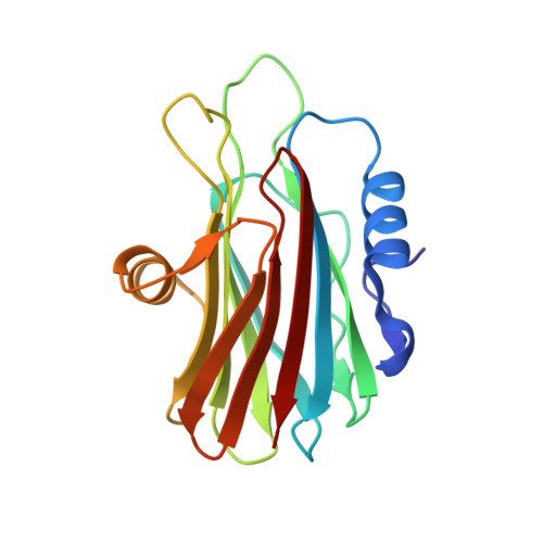
    Sticholysin II (StnII) is a pore-forming protein (PFP) produced by the sea anemone Stichodactyla helianthus. We found out that StnII exists in a monomeric soluble state but forms tetramers in the presence of a lipidic interface. Both structures have been independently determined at 1.7 A and 18 A resolution, respectively, by using X-ray crystallography and electron microscopy of two-dimensional crystals. Besides, the structure of soluble StnII complexed with phosphocholine, determined at 2.4 A resolution, reveals a phospholipid headgroup binding site, which is located in a region with an unusually high abundance of aromatic residues. Fitting of the atomic model into the electron microscopy density envelope suggests that while the beta sandwich structure of the protein remains intact upon oligomerization, the N-terminal region and a flexible and highly basic loop undergo significant conformational changes. These results provide the structural basis for the membrane recognition step of actinoporins and unexpected insights into the oligomerization step.


  • Organizational Affiliation
    • Grupo de Cristalografía Macromolecular y Biología Estructural, Instituto Rocasolano, CSIC, Serrano 119, 28006 Madrid, Spain.

Macromolecule Content 

  • Total Structure Weight: 39.16 kDa 
  • Atom Count: 2,900 
  • Modeled Residue Count: 350 
  • Deposited Residue Count: 350 
  • Unique protein chains: 1

Macromolecules

Find similar proteins by:|  3D Structure
Entity ID: 1
MoleculeChains  Sequence LengthOrganismDetailsImage
STICHOLYSIN II
A, B
175Stichodactyla helianthusMutation(s): 0 
UniProt
Find proteins for P07845 (Stichodactyla helianthus)
Explore P07845 
Go to UniProtKB:  P07845
Entity Groups
Sequence Clusters30% Identity50% Identity70% Identity90% Identity95% Identity100% Identity
UniProt GroupP07845
Sequence Annotations
Expand
Reference Sequence

Experimental Data & Validation

Experimental Data

  • Method: X-RAY DIFFRACTION
  • Resolution: 2.26 Å
  • R-Value Free:  0.269 (Depositor), 0.273 (DCC) 
  • R-Value Work:  0.215 (Depositor), 0.218 (DCC) 
  • R-Value Observed: 0.215 (Depositor) 
Space Group: P 1 21 1
Unit Cell:
Length ( Å )Angle ( ˚ )
a = 32.86α = 90
b = 43.57β = 92.43
c = 120.03γ = 90
Software Package:
Software NamePurpose
CNSrefinement
MOSFLMdata reduction

Structure Validation

View Full Validation Report


Entry History 

Deposition Data

Revision History  (Full details and data files)

  • Version 1.0: 2003-11-13
    Type: Initial release
  • Version 1.1: 2011-05-08
    Changes: Version format compliance
  • Version 1.2: 2011-07-13
    Changes: Version format compliance
  • Version 1.3: 2024-05-08
    Changes: Data collection, Database references, Other