1NYT | pdb_00001nyt

SHIKIMATE DEHYDROGENASE AroE COMPLEXED WITH NADP+


Experimental Data Snapshot

  • Method: X-RAY DIFFRACTION
  • Resolution: 1.50 Å
  • R-Value Free: 
    0.169 (Depositor) 
  • R-Value Work: 
    0.132 (Depositor), 0.164 (DCC) 
  • R-Value Observed: 
    0.136 (Depositor) 

wwPDB Validation 3D Report Full Report

Validation slider image for 1NYT

Ligand Structure Quality Assessment 


This is version 1.4 of the entry. See complete history

Literature

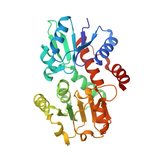
Structures of shikimate dehydrogenase AroE and its Paralog YdiB. A common structural framework for different activities.

Michel, G.Roszak, A.W.Sauve, V.Maclean, J.Matte, A.Coggins, J.R.Cygler, M.Lapthorn, A.J.

(2003) J Biological Chem 278: 19463-19472

  • DOI: https://doi.org/10.1074/jbc.M300794200
  • Primary Citation Related Structures: 
    1NYT, 1O9B

  • PubMed Abstract: 

    Shikimate dehydrogenase catalyzes the fourth step of the shikimate pathway, the essential route for the biosynthesis of aromatic compounds in plants and microorganisms. Absent in metazoans, this pathway is an attractive target for nontoxic herbicides and drugs. Escherichia coli expresses two shikimate dehydrogenase paralogs, the NADP-specific AroE and a putative enzyme YdiB. Here we characterize YdiB as a dual specificity quinate/shikimate dehydrogenase that utilizes either NAD or NADP as a cofactor. Structures of AroE and YdiB with bound cofactors were determined at 1.5 and 2.5 A resolution, respectively. Both enzymes display a similar architecture with two alpha/beta domains separated by a wide cleft. Comparison of their dinucleotide-binding domains reveals the molecular basis for cofactor specificity. Independent molecules display conformational flexibility suggesting that a switch between open and closed conformations occurs upon substrate binding. Sequence analysis and structural comparison led us to propose the catalytic machinery and a model for 3-dehydroshikimate recognition. Furthermore, we discuss the evolutionary and metabolic implications of the presence of two shikimate dehydrogenases in E. coli and other organisms.


  • Organizational Affiliation
    • Biotechnology Research Institute, NRC Macromolecular Structure Group, Montreal, Quebec H4P 2R2, Canada.

Macromolecule Content 

  • Total Structure Weight: 121.96 kDa 
  • Atom Count: 9,929 
  • Modeled Residue Count: 1,081 
  • Deposited Residue Count: 1,084 
  • Unique protein chains: 1

Macromolecules

Find similar proteins by:|  3D Structure
Entity ID: 1
MoleculeChains  Sequence LengthOrganismDetailsImage
Shikimate 5-dehydrogenase
A, B, C, D
271Escherichia coliMutation(s): 0 
Gene Names: AROE
EC: 1.1.1.25
UniProt
Find proteins for P15770 (Escherichia coli (strain K12))
Explore P15770 
Go to UniProtKB:  P15770
Entity Groups
Sequence Clusters30% Identity50% Identity70% Identity90% Identity95% Identity100% Identity
UniProt GroupP15770
Sequence Annotations
Expand
Reference Sequence

Small Molecules

Ligands 3 Unique
IDChains Name / Formula / InChI Key2D Diagram3D Interactions
NAP

Query on NAP


Download:Ideal Coordinates CCD File
I [auth A],
O [auth B],
T [auth C],
W [auth D]
NADP NICOTINAMIDE-ADENINE-DINUCLEOTIDE PHOSPHATE
C21 H28 N7 O17 P3
XJLXINKUBYWONI-NNYOXOHSSA-N
DTV

Query on DTV


Download:Ideal Coordinates CCD File
J [auth A](2S,3S)-1,4-DIMERCAPTOBUTANE-2,3-DIOL
C4 H10 O2 S2
VHJLVAABSRFDPM-QWWZWVQMSA-N
SO4

Query on SO4


Download:Ideal Coordinates CCD File
E [auth A]
F [auth A]
G [auth A]
H [auth A]
K [auth B]
E [auth A],
F [auth A],
G [auth A],
H [auth A],
K [auth B],
L [auth B],
M [auth B],
N [auth B],
P [auth C],
Q [auth C],
R [auth C],
S [auth C],
U [auth D],
V [auth D]
SULFATE ION
O4 S
QAOWNCQODCNURD-UHFFFAOYSA-L

Experimental Data & Validation

Experimental Data

  • Method: X-RAY DIFFRACTION
  • Resolution: 1.50 Å
  • R-Value Free:  0.169 (Depositor) 
  • R-Value Work:  0.132 (Depositor), 0.164 (DCC) 
  • R-Value Observed: 0.136 (Depositor) 
Space Group: C 1 2 1
Unit Cell:
Length ( Å )Angle ( ˚ )
a = 110.524α = 90
b = 140.019β = 122.07
c = 102.712γ = 90
Software Package:
Software NamePurpose
REFMACrefinement
DENZOdata reduction
SCALEPACKdata scaling
CCP4data scaling
MLPHAREphasing
SHELXDphasing
DMphasing
SOLVEphasing

Structure Validation

View Full Validation Report


Ligand Structure Quality Assessment 


Entry History 

Deposition Data

Revision History  (Full details and data files)

  • Version 1.0: 2003-03-04
    Type: Initial release
  • Version 1.1: 2008-04-29
    Changes: Version format compliance
  • Version 1.2: 2011-07-13
    Changes: Atomic model, Version format compliance
  • Version 1.3: 2024-02-14
    Changes: Data collection, Database references, Derived calculations
  • Version 1.4: 2024-03-13
    Changes: Source and taxonomy, Structure summary