1NYL | pdb_00001nyl

Unliganded glutaminyl-tRNA synthetase


Experimental Data Snapshot

  • Method: X-RAY DIFFRACTION
  • Resolution: 2.60 Å
  • R-Value Free: 
    0.327 (Depositor) 
  • R-Value Work: 
    0.251 (Depositor), 0.252 (DCC) 
  • R-Value Observed: 
    0.251 (Depositor) 

wwPDB Validation 3D Report Full Report

Validation slider image for 1NYL

This is version 1.3 of the entry. See complete history

Literature

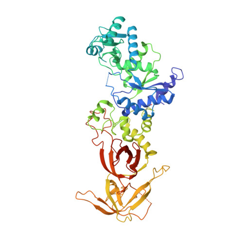
tRNA-Dependent Active Site Assembly in a Class I Aminoacyl-tRNA Synthetase.

Sherlin, L.D.Perona, J.J.

(2003) Structure 11: 591-603

  • DOI: https://doi.org/10.1016/s0969-2126(03)00074-1
  • Primary Citation Related Structures: 
    1NYL

  • PubMed Abstract: 

    The crystal structure of ligand-free E. coli glutaminyl-tRNA synthetase (GlnRS) at 2.4 A resolution shows that substrate binding is essential to construction of a catalytically proficient active site. tRNA binding generates structural changes throughout the enzyme, repositioning key active site peptides that bind glutamine and ATP. The structure gives insight into longstanding questions regarding the tRNA dependence of glutaminyl adenylate formation, the coupling of amino acid and tRNA selectivities, and the roles of specific pathways for transmission of tRNA binding signals to the active site. Comparative analysis of the unliganded and tRNA-bound structures shows, in detail, how flexibility is built into the enzyme architecture and suggests that the induced-fit transitions are a key underlying determinant of both amino acid and tRNA specificity.


  • Organizational Affiliation
    • Department of Chemistry and Biochemistry, University of California, Santa Barbara, Santa Barbara, CA 93106, USA.

Macromolecule Content 

  • Total Structure Weight: 61.92 kDa 
  • Atom Count: 4,240 
  • Modeled Residue Count: 523 
  • Deposited Residue Count: 539 
  • Unique protein chains: 1

Macromolecules

Find similar proteins by:|  3D Structure
Entity ID: 1
MoleculeChains  Sequence LengthOrganismDetailsImage
Glutaminyl-tRNA synthetase539Escherichia coliMutation(s): 0 
Gene Names: glnS
EC: 6.1.1.18
UniProt
Find proteins for P00962 (Escherichia coli (strain K12))
Explore P00962 
Go to UniProtKB:  P00962
Entity Groups
Sequence Clusters30% Identity50% Identity70% Identity90% Identity95% Identity100% Identity
UniProt GroupP00962
Sequence Annotations
Expand
Reference Sequence

Experimental Data & Validation

Experimental Data

  • Method: X-RAY DIFFRACTION
  • Resolution: 2.60 Å
  • R-Value Free:  0.327 (Depositor) 
  • R-Value Work:  0.251 (Depositor), 0.252 (DCC) 
  • R-Value Observed: 0.251 (Depositor) 
Space Group: P 21 21 21
Unit Cell:
Length ( Å )Angle ( ˚ )
a = 41.4α = 90
b = 64.12β = 90
c = 208.88γ = 90
Software Package:
Software NamePurpose
CNSrefinement

Structure Validation

View Full Validation Report


Entry History 

Deposition Data

Revision History  (Full details and data files)

  • Version 1.0: 2003-02-25
    Type: Initial release
  • Version 1.1: 2008-04-29
    Changes: Version format compliance
  • Version 1.2: 2011-07-13
    Changes: Version format compliance
  • Version 1.3: 2024-11-20
    Changes: Data collection, Database references, Structure summary