1NVT | pdb_00001nvt

Crystal structure of Shikimate Dehydrogenase (AROE or MJ1084) in complex with NADP+


Experimental Data Snapshot

  • Method: X-RAY DIFFRACTION
  • Resolution: 2.35 Å
  • R-Value Free: 
    0.260 (Depositor), 0.251 (DCC) 
  • R-Value Work: 
    0.224 (Depositor), 0.217 (DCC) 
  • R-Value Observed: 
    0.224 (Depositor) 

wwPDB Validation 3D Report Full Report

Validation slider image for 1NVT

Ligand Structure Quality Assessment 


This is version 1.4 of the entry. See complete history

Literature

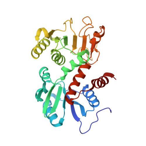
Crystal structure of shikimate 5-dehydrogenase (SDH) bound to NADP: insights into function and evolution

Padyana, A.K.Burley, S.K.

(2003) Structure 11: 1005-1013

  • DOI: https://doi.org/10.1016/s0969-2126(03)00159-x
  • Primary Citation Related Structures: 
    1NVT

  • PubMed Abstract: 

    The crystal structure of Methanococcus jannaschii shikimate 5-dehydrogenase (MjSDH) bound to the cofactor nicotinamide adenine dinucleotide phosphate (NADP) has been determined at 2.35 A resolution. Shikimate 5-dehydrogenase (SDH) is responsible for NADP-dependent catalysis of the fourth step in shikimate biosynthesis, which is essential for aromatic amino acid metabolism in bacteria, microbial eukaryotes, and plants. The structure of MjSDH is a compact alpha/beta sandwich with two distinct domains, responsible for binding substrate and the NADP cofactor, respectively. A phylogenetically conserved deep cleft on the protein surface corresponds to the enzyme active site. The structure reveals a topologically new domain fold within the N-terminal segment of the polypeptide chain, which binds substrate and supports dimerization. Insights gained from homology modeling and sequence/structure comparisons suggest that the SDHs represent a unique class of dehydrogenases. The structure provides a framework for further investigation to discover and develop novel inhibitors targeting this essential enzyme.


  • Organizational Affiliation
    • Laboratories of Molecular Biophysics, The Rockefeller University, 1230 York Avenue, New York, NY 10021, USA.

Macromolecule Content 

  • Total Structure Weight: 64.27 kDa 
  • Atom Count: 4,637 
  • Modeled Residue Count: 574 
  • Deposited Residue Count: 574 
  • Unique protein chains: 1

Macromolecules

Find similar proteins by:|  3D Structure
Entity ID: 1
MoleculeChains  Sequence LengthOrganismDetailsImage
Shikimate 5'-dehydrogenase
A, B
287Methanocaldococcus jannaschiiMutation(s): 0 
Gene Names: aroE
EC: 1.1.1.25
UniProt
Find proteins for Q58484 (Methanocaldococcus jannaschii (strain ATCC 43067 / DSM 2661 / JAL-1 / JCM 10045 / NBRC 100440))
Explore Q58484 
Go to UniProtKB:  Q58484
Entity Groups
Sequence Clusters30% Identity50% Identity70% Identity90% Identity95% Identity100% Identity
UniProt GroupQ58484
Sequence Annotations
Expand
Reference Sequence

Experimental Data & Validation

Experimental Data

  • Method: X-RAY DIFFRACTION
  • Resolution: 2.35 Å
  • R-Value Free:  0.260 (Depositor), 0.251 (DCC) 
  • R-Value Work:  0.224 (Depositor), 0.217 (DCC) 
  • R-Value Observed: 0.224 (Depositor) 
Space Group: P 21 21 21
Unit Cell:
Length ( Å )Angle ( ˚ )
a = 74.65α = 90
b = 75.62β = 90
c = 118.79γ = 90
Software Package:
Software NamePurpose
CNSrefinement
DENZOdata reduction
SCALEPACKdata scaling
SHELXDphasing
MLPHAREphasing
RESOLVEphasing
MAIDphasing

Structure Validation

View Full Validation Report


Ligand Structure Quality Assessment 


Entry History 

Deposition Data

Revision History  (Full details and data files)

  • Version 1.0: 2003-03-25
    Type: Initial release
  • Version 1.1: 2008-04-29
    Changes: Version format compliance
  • Version 1.2: 2011-07-13
    Changes: Version format compliance
  • Version 1.3: 2021-02-03
    Changes: Database references, Derived calculations, Structure summary
  • Version 1.4: 2024-02-14
    Changes: Data collection, Database references