1NSN | pdb_00001nsn

THE CRYSTAL STRUCTURE OF ANTIBODY N10-STAPHYLOCOCCAL NUCLEASE COMPLEX AT 2.9 ANGSTROMS RESOLUTION


Experimental Data Snapshot

  • Method: X-RAY DIFFRACTION
  • Resolution: 2.80 Å
  • R-Value Work: 
    0.195 (Depositor), 0.195 (DCC) 
  • R-Value Observed: 
    0.195 (Depositor) 

wwPDB Validation 3D Report Full Report

Validation slider image for 1NSN

This is version 1.4 of the entry. See complete history

Literature

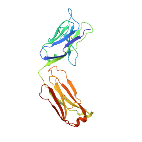
The crystal structure of the antibody N10-staphylococcal nuclease complex at 2.9 A resolution.

Bossart-Whitaker, P.Chang, C.Y.Novotny, J.Benjamin, D.C.Sheriff, S.

(1995) J Mol Biology 253: 559-575

  • DOI: https://doi.org/10.1006/jmbi.1995.0573
  • Primary Citation Related Structures: 
    1NSN

  • PubMed Abstract: 

    The three-dimensional structure of the antibody N10 Fab fragment complexed with staphylococcal nuclease (SNase) has been determined to 2.9 A resolution. Eighteen residues from six complementarity-determining regions (CDR) recognize an epitope of five distinct SNase segments with a total of 17 residues. The overall shape of the antibody-antigen interface is U-shaped rather than the more or less rectangular interface seen in other antibody-protein antigen interfaces. Despite the U-shaped interface, the amount of surface buried in the complex, 828 A2 for SNase and 793 A2 for N10, is typical of antibody-protein antigen complexes. Contributing to the shape of the interface is the shortest antibody heavy chain-CDR3 loop reported to date, which probably allows access of bulk solvent in the center of the "U" interface. Another unusual feature of the N10 antibody is the 15 residue antibody light chain-CDR1, a length seen in only three other reported antibodies. Antibody light chain-CDR1 displays a previously unobserved conformation in its distal portion. Finally, although some of the movement observed in the antibody-bound SNase may be due to crystal contacts, it is clear that some side-chain rearrangements are the result of antigen-antibody interaction.


  • Organizational Affiliation
    • Bristol-Myers Squibb Pharmaceutical Research Institute, Princeton, NJ 08543-4000, USA.

Macromolecule Content 

  • Total Structure Weight: 63.61 kDa 
  • Atom Count: 4,388 
  • Modeled Residue Count: 565 
  • Deposited Residue Count: 577 
  • Unique protein chains: 3

Macromolecules

Find similar proteins by:|  3D Structure
Entity ID: 1
MoleculeChains  Sequence LengthOrganismDetailsImage
IGG FAB (IGG1, KAPPA)A [auth L]218Mus musculusMutation(s): 0 
Find similar proteins by:|  3D Structure
Entity ID: 2
MoleculeChains  Sequence LengthOrganismDetailsImage
IGG FAB (IGG1, KAPPA)B [auth H]210Mus musculusMutation(s): 0 
Find similar proteins by:|  3D Structure
Entity ID: 3
MoleculeChains  Sequence LengthOrganismDetailsImage
STAPHYLOCOCCAL NUCLEASEC [auth S]149Staphylococcus aureusMutation(s): 0 
Gene Names: NUCLEASE
EC: 3.1.31.1
UniProt
Find proteins for P00644 (Staphylococcus aureus)
Explore P00644 
Go to UniProtKB:  P00644
Entity Groups
Sequence Clusters30% Identity50% Identity70% Identity90% Identity95% Identity100% Identity
UniProt GroupP00644
Sequence Annotations
Expand
Reference Sequence

Experimental Data & Validation

Experimental Data

  • Method: X-RAY DIFFRACTION
  • Resolution: 2.80 Å
  • R-Value Work:  0.195 (Depositor), 0.195 (DCC) 
  • R-Value Observed: 0.195 (Depositor) 
Space Group: C 1 2 1
Unit Cell:
Length ( Å )Angle ( ˚ )
a = 234.7α = 90
b = 43.5β = 106.4
c = 74.4γ = 90
Software Package:
Software NamePurpose
XENGENdata collection
X-PLORmodel building
X-PLORrefinement
XENGENdata reduction
X-PLORphasing

Structure Validation

View Full Validation Report


Entry History 

Deposition Data

Revision History  (Full details and data files)

  • Version 1.0: 1995-09-15
    Type: Initial release
  • Version 1.1: 2008-03-24
    Changes: Version format compliance
  • Version 1.2: 2011-07-13
    Changes: Version format compliance
  • Version 1.3: 2024-06-05
    Changes: Data collection, Database references, Other
  • Version 1.4: 2024-11-20
    Changes: Structure summary