1NQD

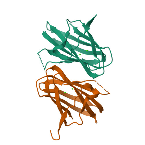
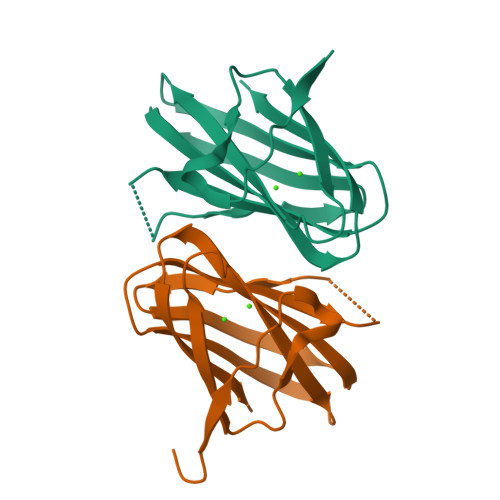
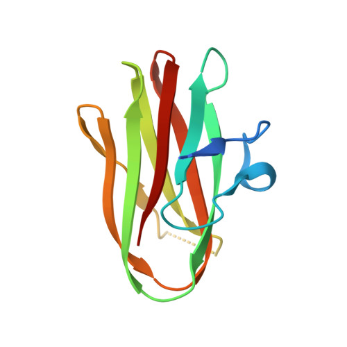
CRYSTAL STRUCTURE OF CLOSTRIDIUM HISTOLYTICUM COLG COLLAGENASE COLLAGEN-BINDING DOMAIN 3B AT 1.65 ANGSTROM RESOLUTION IN PRESENCE OF CALCIUM


Experimental Data Snapshot

  • Method: X-RAY DIFFRACTION
  • Resolution: 1.65 Å
  • R-Value Free: 0.254 
  • R-Value Work: 0.185 
  • R-Value Observed: 0.191 

Starting Model: experimental
View more details

wwPDB Validation   3D Report Full Report


This is version 1.5 of the entry. See complete history


Literature

A Bacterial Collagen-Binding Domain with Novel Calcium-Binding Motif Controls Domain Orientation

Wilson, J.J.Matsushita, O.Okabe, A.Sakon, J.

(2003) EMBO J 22: 1743-1752

  • DOI: https://doi.org/10.1093/emboj/cdg172
  • Primary Citation of Related Structures:  
    1NQD, 1NQJ

  • PubMed Abstract: 

    The crystal structure of a collagen-binding domain (CBD) with an N-terminal domain linker from Clostridium histolyticum class I collagenase was determined at 1.00 A resolution in the absence of calcium (1NQJ) and at 1.65 A resolution in the presence of calcium (1NQD). The mature enzyme is composed of four domains: a metalloprotease domain, a spacing domain and two CBDs. A 12-residue-long linker is found at the N-terminus of each CBD. In the absence of calcium, the CBD reveals a beta-sheet sandwich fold with the linker adopting an alpha-helix. The addition of calcium unwinds the linker and anchors it to the distal side of the sandwich as a new beta-strand. The conformational change of the linker upon calcium binding is confirmed by changes in the Stokes and hydrodynamic radii as measured by size exclusion chromatography and by dynamic light scattering with and without calcium. Furthermore, extensive mutagenesis of conserved surface residues and collagen-binding studies allow us to identify the collagen-binding surface of the protein and propose likely collagen-protein binding models.


  • Organizational Affiliation

    Department of Chemistry and Biochemistry, University of Arkansas, Fayetteville, AR 72701, USA.


Macromolecules
Find similar proteins by:  (by identity cutoff)  |  3D Structure
Entity ID: 1
MoleculeChains Sequence LengthOrganismDetailsImage
class 1 collagenase
A, B
122Hathewaya histolyticaMutation(s): 0 
Gene Names: ColG
EC: 3.4.24.3
UniProt
Find proteins for Q9X721 (Hathewaya histolytica)
Explore Q9X721 
Go to UniProtKB:  Q9X721
Entity Groups  
Sequence Clusters30% Identity50% Identity70% Identity90% Identity95% Identity100% Identity
UniProt GroupQ9X721
Sequence Annotations
Expand
  • Reference Sequence
Experimental Data & Validation

Experimental Data

  • Method: X-RAY DIFFRACTION
  • Resolution: 1.65 Å
  • R-Value Free: 0.254 
  • R-Value Work: 0.185 
  • R-Value Observed: 0.191 
  • Space Group: P 1 21 1
Unit Cell:
Length ( Å )Angle ( ˚ )
a = 40.867α = 90
b = 59.401β = 98.73
c = 48.572γ = 90
Software Package:
Software NamePurpose
SHELXL-97refinement
HKL-2000data reduction
CNSrefinement
SHELXmodel building
HKL-2000data collection
HKL-2000data scaling
CNSphasing

Structure Validation

View Full Validation Report


Entry History 

Deposition Data

Revision History  (Full details and data files)

  • Version 1.0: 2003-04-15
    Type: Initial release
  • Version 1.1: 2008-04-29
    Changes: Version format compliance
  • Version 1.2: 2011-07-13
    Changes: Version format compliance
  • Version 1.3: 2019-07-24
    Changes: Data collection, Refinement description
  • Version 1.4: 2024-02-14
    Changes: Data collection, Database references, Derived calculations
  • Version 1.5: 2024-04-03
    Changes: Refinement description