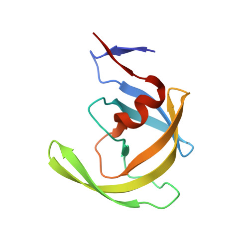
Design, synthesis, and biological evaluation of monopyrrolinone-based HIV-1 protease inhibitors.
Smith III, A.B., Cantin, L.D., Pasternak, A., Guise-Zawacki, L., Yao, W., Charnley, A.K., Barbosa, J., Sprengeler, P.A., Hirschmann, R., Munshi, S., Olsen, D.B., Schleif, W.A., Kuo, L.C.(2003) J Med Chem 46: 1831-1844
- PubMed: 12723947
- DOI: https://doi.org/10.1021/jm0204587
- Primary Citation of Related Structures:
1NPV, 1NPW - PubMed Abstract:
The design, synthesis, and biological evaluation of a series of HIV-1 protease inhibitors [(-)-6, (-)-7, (-)-23, (+)-24] based upon the 3,5,5-trisubstituted pyrrolin-4-one scaffold is described. Use of a monopyrrolinone scaffold leads to inhibitors with improved cellular transport properties relative to the earlier inhibitors based on bispyrrolinones and their peptide counterparts. The most potent inhibitor (-)-7 displayed 13% oral bioavailability in dogs. X-ray structure analysis of the monopyrrolinone compounds cocrystallized with the wild-type HIV-1 protease provided valuable information on the interactions between the inhibitors and the HIV-1 enzyme. In each case, the inhibitors assumed similar orientations for the P2'-P1 substituents, along with an unexpected hydrogen bond of the pyrrolinone NH with Asp225. Interactions with the S2 pocket, however, were not optimal, as illustrated by the inclusion of a water molecule in two of the three inhibitor-enzyme complexes. Efforts to increase affinity by displacing the water molecule with second and third generation inhibitors did not prove successful. Lack of success with this venture is a testament to the difficulty of accurately predicting the many variables that influence and build binding affinity. Comparison of the inhibitor positions in three complexes with that of Indinavir revealed displacements of the protease backbones in the enzyme flap region, accompanied by variations in hydrogen bonding to accommodate the monopyrrolinone ring. The binding orientation of the pyrrolinone-based inhibitors may explain their sustained efficacy against mutant strains of the HIV-1 protease enzyme as compared to Indinavir.
Organizational Affiliation:
Department of Chemistry, University of Pennsylvania, 231 South 34th Street, Philadelphia, Pennsylvania 19104, USA. smithab@sas.upenn.edu


