1NOX | pdb_00001nox

NADH OXIDASE FROM THERMUS THERMOPHILUS


Experimental Data Snapshot

  • Method: X-RAY DIFFRACTION
  • Resolution: 1.59 Å
  • R-Value Free: 
    0.204 (Depositor) 
  • R-Value Work: 
    0.182 (Depositor), 0.176 (DCC) 
  • R-Value Observed: 
    0.190 (Depositor) 

Starting Model: experimental
View more details

wwPDB Validation 3D Report Full Report

Validation slider image for 1NOX

Ligand Structure Quality Assessment 


This is version 1.4 of the entry. See complete history

Literature

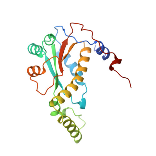
Crystal structure of NADH oxidase from Thermus thermophilus.

Hecht, H.J.Erdmann, H.Park, H.J.Sprinzl, M.Schmid, R.D.

(1995) Nat Struct Biol 2: 1109-1114

  • DOI: https://doi.org/10.1038/nsb1295-1109
  • Primary Citation Related Structures: 
    1NOX

  • PubMed Abstract: 

    The crystal structures of the flavin adenine dinucleotide (FAD) and flavin mononucleotide (FMN) containing isoforms of NADH oxidase from Thermus thermophilus have been determined by isomorphous and molecular replacement and refined to 2.3 A and 1.6 A resolution with R-values of 18.5% and 18.6% respectively. The structure of the homodimeric enzyme consists of a central 4-stranded antiparallel beta-sheet covered by helices, a more flexible domain formed by two helices, and a C-terminal excursion connecting the subunits. The active sites are located in a deep cleft between the subunits. The binding site of the flavin cofactor lacks the common nucleotide binding fold and is different from the FMN binding site found in flavodoxins.


  • Organizational Affiliation
    • Department of Molecular Structure Research, GBF (Gesellschaft fr Biotechnologische, Forschung, Braunschweig, Germany.

Macromolecule Content 

  • Total Structure Weight: 23.24 kDa 
  • Atom Count: 1,695 
  • Modeled Residue Count: 200 
  • Deposited Residue Count: 205 
  • Unique protein chains: 1

Macromolecules

Find similar proteins by:|  3D Structure
Entity ID: 1
MoleculeChains  Sequence LengthOrganismDetailsImage
NADH OXIDASE205Thermus thermophilus HB8Mutation(s): 0 
EC: 1.6.99.3 (PDB Primary Data), 7.1.1.2 (UniProt)
UniProt
Find proteins for Q60049 (Thermus thermophilus (strain ATCC 27634 / DSM 579 / HB8))
Explore Q60049 
Go to UniProtKB:  Q60049
Entity Groups
Sequence Clusters30% Identity50% Identity70% Identity90% Identity95% Identity100% Identity
UniProt GroupQ60049
Sequence Annotations
Expand
Reference Sequence

Small Molecules

Ligands 1 Unique
IDChains Name / Formula / InChI Key2D Diagram3D Interactions
FMN

Query on FMN


Download:Ideal Coordinates CCD File
B [auth A]FLAVIN MONONUCLEOTIDE
C17 H21 N4 O9 P
FVTCRASFADXXNN-SCRDCRAPSA-N

Experimental Data & Validation

Experimental Data

  • Method: X-RAY DIFFRACTION
  • Resolution: 1.59 Å
  • R-Value Free:  0.204 (Depositor) 
  • R-Value Work:  0.182 (Depositor), 0.176 (DCC) 
  • R-Value Observed: 0.190 (Depositor) 
Space Group: P 21 21 2
Unit Cell:
Length ( Å )Angle ( ˚ )
a = 62.53α = 90
b = 60.6β = 90
c = 56.66γ = 90
Software Package:
Software NamePurpose
X-PLORmodel building
REFMACrefinement
X-PLORrefinement
MOSFLMdata reduction
CCP4data scaling
SCALAdata scaling
X-PLORphasing

Structure Validation

View Full Validation Report


Ligand Structure Quality Assessment 


Entry History 

Deposition Data

Revision History  (Full details and data files)

  • Version 1.0: 1997-03-12
    Type: Initial release
  • Version 1.1: 2008-03-24
    Changes: Version format compliance
  • Version 1.2: 2011-07-13
    Changes: Derived calculations, Source and taxonomy, Version format compliance
  • Version 1.3: 2024-02-14
    Changes: Data collection, Database references, Derived calculations, Other
  • Version 1.4: 2024-04-03
    Changes: Refinement description