1NOD | pdb_00001nod

MURINE INDUCIBLE NITRIC OXIDE SYNTHASE OXYGENASE DIMER (DELTA 65) WITH TETRAHYDROBIOPTERIN AND SUBSTRATE L-ARGININE


Experimental Data Snapshot

  • Method: X-RAY DIFFRACTION
  • Resolution: 2.60 Å
  • R-Value Free: 
    0.288 (Depositor) 
  • R-Value Work: 
    0.229 (Depositor) 
  • R-Value Observed: 
    0.229 (Depositor) 

Starting Model: experimental
View more details

wwPDB Validation 3D Report Full Report

Validation slider image for 1NOD

This is version 1.5 of the entry. See complete history

Literature

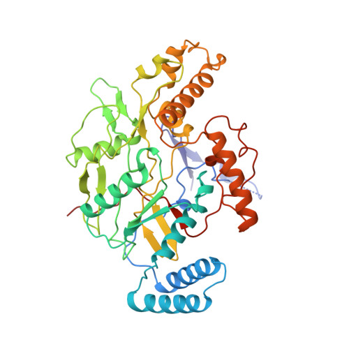
Structure of nitric oxide synthase oxygenase dimer with pterin and substrate.

Crane, B.R.Arvai, A.S.Ghosh, D.K.Wu, C.Getzoff, E.D.Stuehr, D.J.Tainer, J.A.

(1998) Science 279: 2121-2126

  • DOI: https://doi.org/10.1126/science.279.5359.2121
  • Primary Citation Related Structures: 
    1NOD, 2NOD, 3NOD

  • PubMed Abstract: 

    Crystal structures of the murine cytokine-inducible nitric oxide synthase oxygenase dimer with active-center water molecules, the substrate L-arginine (L-Arg), or product analog thiocitrulline reveal how dimerization, cofactor tetrahydrobiopterin, and L-Arg binding complete the catalytic center for synthesis of the essential biological signal and cytotoxin nitric oxide. Pterin binding refolds the central interface region, recruits new structural elements, creates a 30 angstrom deep active-center channel, and causes a 35 degrees helical tilt to expose a heme edge and the adjacent residue tryptophan-366 for likely reductase domain interactions and caveolin inhibition. Heme propionate interactions with pterin and L-Arg suggest that pterin has electronic influences on heme-bound oxygen. L-Arginine binds to glutamic acid-371 and stacks with heme in an otherwise hydrophobic pocket to aid activation of heme-bound oxygen by direct proton donation and thereby differentiate the two chemical steps of nitric oxide synthesis.


  • Organizational Affiliation
    • Department of Molecular Biology and Skaggs Institute for Chemical Biology, The Scripps Research Institute, La Jolla, CA 92037, USA.

Macromolecule Content 

  • Total Structure Weight: 100.16 kDa 
  • Atom Count: 7,208 
  • Modeled Residue Count: 827 
  • Deposited Residue Count: 846 
  • Unique protein chains: 1

Macromolecules

Find similar proteins by:|  3D Structure
Entity ID: 1
MoleculeChains  Sequence LengthOrganismDetailsImage
NITRIC OXIDE SYNTHASE
A, B
423Mus musculusMutation(s): 0 
EC: 1.14.13.39
UniProt & NIH Common Fund Data Resources
Find proteins for P29477 (Mus musculus)
Explore P29477 
Go to UniProtKB:  P29477
IMPC:  MGI:97361
Entity Groups
Sequence Clusters30% Identity50% Identity70% Identity90% Identity95% Identity100% Identity
UniProt GroupP29477
Sequence Annotations
Expand
Reference Sequence

Small Molecules

Ligands 4 Unique
IDChains Name / Formula / InChI Key2D Diagram3D Interactions
HEM

Query on HEM


Download:Ideal Coordinates CCD File
E [auth A],
H [auth B]
PROTOPORPHYRIN IX CONTAINING FE
C34 H32 Fe N4 O4
KABFMIBPWCXCRK-RGGAHWMASA-L
H4B

Query on H4B


Download:Ideal Coordinates CCD File
F [auth A],
I [auth B]
5,6,7,8-TETRAHYDROBIOPTERIN
C9 H15 N5 O3
FNKQXYHWGSIFBK-RPDRRWSUSA-N
ARG

Query on ARG


Download:Ideal Coordinates CCD File
D [auth A],
G [auth B]
ARGININE
C6 H15 N4 O2
ODKSFYDXXFIFQN-BYPYZUCNSA-O
SO4

Query on SO4


Download:Ideal Coordinates CCD File
C [auth A]SULFATE ION
O4 S
QAOWNCQODCNURD-UHFFFAOYSA-L

Experimental Data & Validation

Experimental Data

  • Method: X-RAY DIFFRACTION
  • Resolution: 2.60 Å
  • R-Value Free:  0.288 (Depositor) 
  • R-Value Work:  0.229 (Depositor) 
  • R-Value Observed: 0.229 (Depositor) 
Space Group: P 61 2 2
Unit Cell:
Length ( Å )Angle ( ˚ )
a = 213α = 90
b = 213β = 90
c = 114.2γ = 120
Software Package:
Software NamePurpose
DENZOdata reduction
SCALEPACKdata scaling
X-PLORmodel building
X-PLORrefinement
X-PLORphasing

Structure Validation

View Full Validation Report


Entry History 

Deposition Data

Revision History  (Full details and data files)

  • Version 1.0: 1999-03-23
    Type: Initial release
  • Version 1.1: 2008-03-24
    Changes: Version format compliance
  • Version 1.2: 2011-07-13
    Changes: Derived calculations, Version format compliance
  • Version 1.3: 2011-11-16
    Changes: Atomic model
  • Version 1.4: 2023-08-09
    Changes: Database references, Derived calculations, Refinement description
  • Version 1.5: 2024-11-13
    Changes: Data collection, Structure summary