1NM8 | pdb_00001nm8

Structure of Human Carnitine Acetyltransferase: Molecular Basis for Fatty Acyl Transfer


Experimental Data Snapshot

  • Method: X-RAY DIFFRACTION
  • Resolution: 1.60 Å
  • R-Value Free: 
    0.232 (Depositor), 0.262 (DCC) 
  • R-Value Work: 
    0.208 (Depositor), 0.241 (DCC) 

wwPDB Validation 3D Report Full Report

Validation slider image for 1NM8

This is version 1.3 of the entry. See complete history

Literature

Structure of Human Carnitine Acetyltransferase. Molecular Basis for Fatty Acyl Transfer

Wu, D.Govindasamy, L.Lian, W.Gu, Y.Kukar, T.Agbandje-McKenna, M.McKenna, R.

(2003) J Biological Chem 278: 13159-13165

  • DOI: https://doi.org/10.1074/jbc.M212356200
  • Primary Citation Related Structures: 
    1NM8

  • PubMed Abstract: 

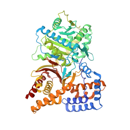
    Carnitine acyltransferases are a family of ubiquitous enzymes that play a pivotal role in cellular energy metabolism. We report here the x-ray structure of human carnitine acetyltransferase to a 1.6-A resolution. This structure reveals a monomeric protein of two equally sized alpha/beta domains. Each domain is shown to have a partially similar fold to other known but oligomeric enzymes that are also involved in group-transfer reactions. The unique monomeric arrangement of the two domains constitutes a central narrow active site tunnel, indicating a likely universal feature for all members of the carnitine acyltransferase family. Superimposition of the substrate complex of a related protein, dihydrolipoyl trans-acetylase, reveals that both substrates localize to the active site tunnel of human carnitine acetyltransferase, suggesting the location of the ligand binding sites for carnitine and coenzyme A. Most significantly, this structure provides critical insights into the molecular basis for fatty acyl chain transfer and a possible common mechanism among a wide range of acyltransferases utilizing a catalytic dyad.


  • Organizational Affiliation
    • Department of Medicinal Chemistry, College of Pharmacy, University of Florida, Gainesville, Florida 32610, USA.

Macromolecule Content 

  • Total Structure Weight: 69.9 kDa 
  • Atom Count: 5,186 
  • Modeled Residue Count: 591 
  • Deposited Residue Count: 616 
  • Unique protein chains: 1

Macromolecules

Find similar proteins by:|  3D Structure
Entity ID: 1
MoleculeChains  Sequence LengthOrganismDetailsImage
Carnitine O-acetyltransferase616Homo sapiensMutation(s): 3 
Gene Names: CRAT OR CAT1
EC: 2.3.1.7 (PDB Primary Data), 2.3.1.137 (UniProt)
Membrane Entity: Yes 
UniProt & NIH Common Fund Data Resources
Find proteins for P43155 (Homo sapiens)
Explore P43155 
Go to UniProtKB:  P43155
PHAROS:  P43155
GTEx:  ENSG00000095321 
Entity Groups
Sequence Clusters30% Identity50% Identity70% Identity90% Identity95% Identity100% Identity
UniProt GroupP43155
Sequence Annotations
Expand
Reference Sequence

Experimental Data & Validation

Experimental Data

  • Method: X-RAY DIFFRACTION
  • Resolution: 1.60 Å
  • R-Value Free:  0.232 (Depositor), 0.262 (DCC) 
  • R-Value Work:  0.208 (Depositor), 0.241 (DCC) 
Space Group: P 21 21 21
Unit Cell:
Length ( Å )Angle ( ˚ )
a = 137.5α = 90
b = 84.5β = 90
c = 57.5γ = 90
Software Package:
Software NamePurpose
DENZOdata reduction
SCALEPACKdata scaling
CNSrefinement
CNSphasing

Structure Validation

View Full Validation Report


Entry History 

Deposition Data

Revision History  (Full details and data files)

  • Version 1.0: 2003-03-11
    Type: Initial release
  • Version 1.1: 2008-04-29
    Changes: Version format compliance
  • Version 1.2: 2011-07-13
    Changes: Version format compliance
  • Version 1.3: 2024-02-14
    Changes: Data collection, Database references