1NM3 | pdb_00001nm3

Crystal structure of Heamophilus influenza hybrid-Prx5


Experimental Data Snapshot

  • Method: X-RAY DIFFRACTION
  • Resolution: 2.80 Å
  • R-Value Free: 
    0.283 (Depositor), 0.280 (DCC) 
  • R-Value Work: 
    0.239 (Depositor), 0.238 (DCC) 
  • R-Value Observed: 
    0.239 (Depositor) 

wwPDB Validation 3D Report Full Report

Validation slider image for 1NM3

This is version 1.3 of the entry. See complete history

Literature

The Tetrameric Structure of Haemophilus influenza Hybrid Prx5 Reveals Interactions between Electron Donor and Acceptor Proteins.

Kim, S.J.Woo, J.R.Hwang, Y.S.Jeong, D.G.Shin, D.H.Kim, K.Ryu, S.E.

(2003) J Biological Chem 278: 10790-10798

  • DOI: https://doi.org/10.1074/jbc.M209553200
  • Primary Citation Related Structures: 
    1NM3

  • PubMed Abstract: 

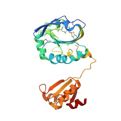
    Cellular redox control is often mediated by oxidation and reduction of cysteine residues in the redox-sensitive proteins, where thioredoxin and glutaredoxin (Grx) play as electron donors for the oxidized proteins. Despite the importance of protein-protein interactions between the electron donor and acceptor proteins, there has been no structural information for the interaction of thioredoxin or Grx with natural target proteins. Here, we present the crystal structure of a novel Haemophilus influenza peroxiredoxin (Prx) hybrid Prx5 determined at 2.8-A resolution. The structure reveals that hybrid Prx5 forms a tightly associated tetramer where active sites of Prx and Grx domains of different monomers interact with each other. The Prx-Grx interface comprises specific charge interactions surrounded by weak interactions, providing insight into the target recognition mechanism of Grx. The tetrameric structure also exhibits a flexible active site and alternative Prx-Grx interactions, which appear to facilitate the electron transfer from Grx to Prx domain. Differences of electron donor binding surfaces in Prx proteins revealed by an analysis based on the structural information explain the electron donor specificities of various Prx proteins.


  • Organizational Affiliation
    • Center for Cellular Switch Protein Structure, Korea Research Institute of Bioscience and Biotechnology, 52 Euh-eun-dong, Yusong-gu, Daejon 305-806, South Korea.

Macromolecule Content 

  • Total Structure Weight: 54.68 kDa 
  • Atom Count: 3,736 
  • Modeled Residue Count: 476 
  • Deposited Residue Count: 482 
  • Unique protein chains: 1

Macromolecules

Find similar proteins by:|  3D Structure
Entity ID: 1
MoleculeChains  Sequence LengthOrganismDetailsImage
Protein HI0572
A, B
241Haemophilus influenzaeMutation(s): 8 
EC: 1.11.1.27
UniProt
Find proteins for P44758 (Haemophilus influenzae (strain ATCC 51907 / DSM 11121 / KW20 / Rd))
Explore P44758 
Go to UniProtKB:  P44758
Entity Groups
Sequence Clusters30% Identity50% Identity70% Identity90% Identity95% Identity100% Identity
UniProt GroupP44758
Sequence Annotations
Expand
Reference Sequence

Experimental Data & Validation

Experimental Data

  • Method: X-RAY DIFFRACTION
  • Resolution: 2.80 Å
  • R-Value Free:  0.283 (Depositor), 0.280 (DCC) 
  • R-Value Work:  0.239 (Depositor), 0.238 (DCC) 
  • R-Value Observed: 0.239 (Depositor) 
Space Group: P 43 21 2
Unit Cell:
Length ( Å )Angle ( ˚ )
a = 72.854α = 90
b = 72.854β = 90
c = 229.633γ = 90
Software Package:
Software NamePurpose
MOSFLMdata reduction
SCALAdata scaling
SHARPphasing
CNSrefinement
CCP4data scaling

Structure Validation

View Full Validation Report


Entry History 

Deposition Data

Revision History  (Full details and data files)

  • Version 1.0: 2003-03-25
    Type: Initial release
  • Version 1.1: 2008-04-29
    Changes: Version format compliance
  • Version 1.2: 2011-07-13
    Changes: Derived calculations, Version format compliance
  • Version 1.3: 2024-11-20
    Changes: Data collection, Database references, Derived calculations, Structure summary