1NIW | pdb_00001niw

Crystal structure of endothelial nitric oxide synthase peptide bound to calmodulin


Experimental Data Snapshot

  • Method: X-RAY DIFFRACTION
  • Resolution: 2.05 Å
  • R-Value Free: 
    0.249 (Depositor), 0.248 (DCC) 
  • R-Value Work: 
    0.223 (Depositor), 0.222 (DCC) 
  • R-Value Observed: 
    0.223 (Depositor) 

wwPDB Validation 3D Report Full Report

Validation slider image for 1NIW

This is version 1.3 of the entry. See complete history

Literature

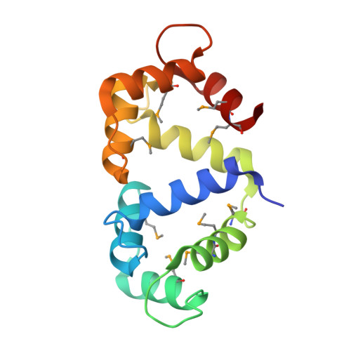
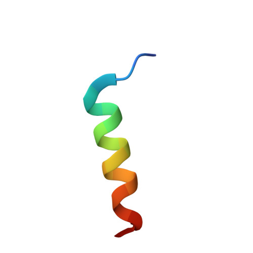
Structural basis for endothelial nitric oxide synthase binding to calmodulin

Aoyagi, M.Arvai, A.S.Tainer, J.A.Getzoff, E.D.

(2003) EMBO J 22: 766-775

  • DOI: https://doi.org/10.1093/emboj/cdg078
  • Primary Citation Related Structures: 
    1NIW

  • PubMed Abstract: 

    The enzyme nitric oxide synthase (NOS) is exquisitely regulated in vivo by the Ca(2+) sensor protein calmodulin (CaM) to control production of NO, a key signaling molecule and cytotoxin. The differential activation of NOS isozymes by CaM has remained enigmatic, despite extensive research. Here, the crystallographic structure of Ca(2+)-loaded CaM bound to a 20 residue peptide comprising the endothelial NOS (eNOS) CaM-binding region establishes their individual conformations and intermolecular interactions, and suggests the basis for isozyme-specific differences. The alpha-helical eNOS peptide binds in an antiparallel orientation to CaM through extensive hydrophobic interactions. Unique NOS interactions occur with: (i). the CaM flexible central linker, explaining its importance in NOS activation; and (ii). the CaM C-terminus, explaining the NOS-specific requirement for a bulky, hydrophobic residue at position 144. This binding mode expands mechanisms for CaM-mediated activation, explains eNOS deactivation by Thr495 phosphorylation, and implicates specific hydrophobic residues in the Ca(2+) independence of inducible NOS.


  • Organizational Affiliation
    • Department of Molecular Biology and The Skaggs Institute for Chemical Biology, The Scripps Research Institute, La Jolla, CA 92037, USA.

Macromolecule Content 

  • Total Structure Weight: 78.29 kDa 
  • Atom Count: 5,460 
  • Modeled Residue Count: 656 
  • Deposited Residue Count: 672 
  • Unique protein chains: 2

Macromolecules

Find similar proteins by:|  3D Structure
Entity ID: 1
MoleculeChains  Sequence LengthOrganismDetailsImage
calmodulin
A, C, E, G
148Rattus norvegicusMutation(s): 9 
UniProt
Find proteins for P0DP29 (Rattus norvegicus)
Explore P0DP29 
Go to UniProtKB:  P0DP29
Entity Groups
Sequence Clusters30% Identity50% Identity70% Identity90% Identity95% Identity100% Identity
UniProt GroupP0DP29
Sequence Annotations
Expand
Reference Sequence
Find similar proteins by:  Sequence   |   3D Structure  
Entity ID: 2
MoleculeChains  Sequence LengthOrganismDetailsImage
Nitric-oxide synthase, endothelial
B, D, F, H
20N/AMutation(s): 0 
EC: 1.14.13.39
UniProt & NIH Common Fund Data Resources
Find proteins for P29474 (Homo sapiens)
Explore P29474 
Go to UniProtKB:  P29474
PHAROS:  P29474
GTEx:  ENSG00000164867 
Entity Groups
Sequence Clusters30% Identity50% Identity70% Identity90% Identity95% Identity100% Identity
UniProt GroupP29474
Sequence Annotations
Expand
Reference Sequence

Small Molecules

Ligands 3 Unique
IDChains Name / Formula / InChI Key2D Diagram3D Interactions
SO4

Query on SO4


Download:Ideal Coordinates CCD File
M [auth A]SULFATE ION
O4 S
QAOWNCQODCNURD-UHFFFAOYSA-L
EDO

Query on EDO


Download:Ideal Coordinates CCD File
R [auth C],
S [auth C],
X [auth E],
Y [auth E]
1,2-ETHANEDIOL
C2 H6 O2
LYCAIKOWRPUZTN-UHFFFAOYSA-N
CA

Query on CA


Download:Ideal Coordinates CCD File
AA [auth G]
BA [auth G]
CA [auth G]
I [auth A]
J [auth A]
AA [auth G],
BA [auth G],
CA [auth G],
I [auth A],
J [auth A],
K [auth A],
L [auth A],
N [auth C],
O [auth C],
P [auth C],
Q [auth C],
T [auth E],
U [auth E],
V [auth E],
W [auth E],
Z [auth G]
CALCIUM ION
Ca
BHPQYMZQTOCNFJ-UHFFFAOYSA-N
Modified Residues  1 Unique
IDChains TypeFormula2D DiagramParent
MSE
Query on MSE
A, C, E, G
L-PEPTIDE LINKINGC5 H11 N O2 SeMET

Experimental Data & Validation

Experimental Data

  • Method: X-RAY DIFFRACTION
  • Resolution: 2.05 Å
  • R-Value Free:  0.249 (Depositor), 0.248 (DCC) 
  • R-Value Work:  0.223 (Depositor), 0.222 (DCC) 
  • R-Value Observed: 0.223 (Depositor) 
Space Group: P 1 21 1
Unit Cell:
Length ( Å )Angle ( ˚ )
a = 68.521α = 90
b = 73.952β = 111.4
c = 71.142γ = 90
Software Package:
Software NamePurpose
CNSrefinement
DENZOdata reduction
SCALEPACKdata scaling
SOLVEphasing
SHARPphasing

Structure Validation

View Full Validation Report


Entry History 

Deposition Data

Revision History  (Full details and data files)

  • Version 1.0: 2003-02-18
    Type: Initial release
  • Version 1.1: 2008-04-29
    Changes: Version format compliance
  • Version 1.2: 2011-07-13
    Changes: Version format compliance
  • Version 1.3: 2024-11-13
    Changes: Data collection, Database references, Derived calculations, Structure summary