1NGK | pdb_00001ngk

Crystallographic Structure of Mycobacterium tuberculosis Hemoglobin O


Experimental Data Snapshot

  • Method: X-RAY DIFFRACTION
  • Resolution: 2.11 Å
  • R-Value Free: 
    0.226 (Depositor), 0.226 (DCC) 
  • R-Value Work: 
    0.186 (Depositor), 0.185 (DCC) 
  • R-Value Observed: 
    0.188 (Depositor) 

wwPDB Validation 3D Report Full Report

Validation slider image for 1NGK

Ligand Structure Quality Assessment 


This is version 1.3 of the entry. See complete history

Literature

A TyrCD1/TrpG8 hydrogen bond network and a TyrB10-TyrCD1 covalent link shape the heme distal site of Mycobacterium tuberculosis hemoglobin O

Milani, M.Savard, P.-Y.Oullet, H.Ascenzi, P.Guertin, M.Bolognesi, M.

(2003) Proc Natl Acad Sci U S A 100: 5766-5771

  • DOI: https://doi.org/10.1073/pnas.1037676100
  • Primary Citation Related Structures: 
    1NGK

  • PubMed Abstract: 

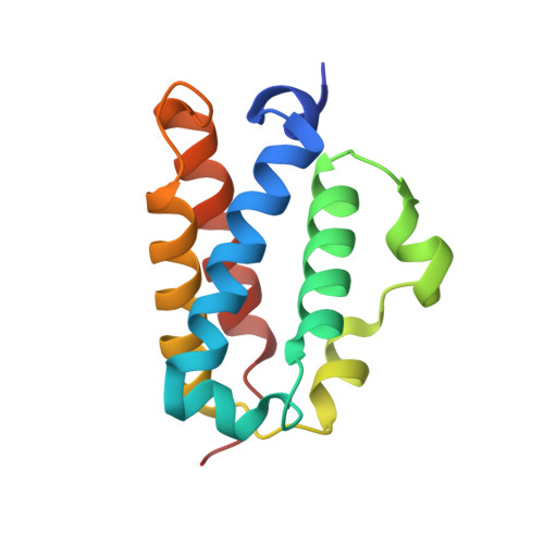
    Truncated hemoglobins (Hbs) are small hemoproteins, identified in microorganisms and in some plants, forming a separate cluster within the Hb superfamily. Two distantly related truncated Hbs, trHbN and trHbO, are expressed at different developmental stages in Mycobacterium tuberculosis. Sequence analysis shows that the two proteins share 18% amino acid identities and belong to different groups within the truncated Hb cluster. Although a specific defense role against nitrosative stress has been ascribed to trHbN (expressed during the Mycobacterium stationary phase), no clear functions have been recognized for trHbO, which is expressed throughout the Mycobacterium growth phase. The 2.1-A crystal structure of M. tuberculosis cyano-met trHbO shows that the protein assembles in a compact dodecamer. Six of the dodecamer subunits are characterized by a double conformation for their CD regions and, most notably, by a covalent bond linking the phenolic O atom of TyrB10 to the aromatic ring of TyrCD1, in the heme distal cavity. All 12 subunits display a cyanide ion bound to the heme Fe atom, stabilized by a tight hydrogen-bonded network based on the (globin very rare) TyrCD1 and TrpG8 residues. The small apolar AlaE7 residue leaves room for ligand access to the heme distal site through the conventional "E7 path," as proposed for myoglobin. Different from trHbN, where a 20-A protein matrix tunnel is held to sustain ligand diffusion to an otherwise inaccessible heme distal site, the topologically related region in trHbO hosts two protein matrix cavities.


  • Organizational Affiliation
    • Department of Physics-National Institute of Physics of Matter, Center for Excellence in Biomedical Research, University of Genoa, Genoa, Italy.

Macromolecule Content 

  • Total Structure Weight: 189.1 kDa 
  • Atom Count: 15,104 
  • Modeled Residue Count: 1,520 
  • Deposited Residue Count: 1,536 
  • Unique protein chains: 1

Macromolecules

Find similar proteins by:|  3D Structure
Entity ID: 1
MoleculeChains  Sequence LengthOrganismDetailsImage
Hemoglobin-like protein HbO
A, B, C, D, E
A, B, C, D, E, F, G, H, I, J, K, L
128Mycobacterium tuberculosisMutation(s): 0 
UniProt
Find proteins for P9WN23 (Mycobacterium tuberculosis (strain ATCC 25618 / H37Rv))
Explore P9WN23 
Go to UniProtKB:  P9WN23
Entity Groups
Sequence Clusters30% Identity50% Identity70% Identity90% Identity95% Identity100% Identity
UniProt GroupP9WN23
Sequence Annotations
Expand
Reference Sequence

Small Molecules

Ligands 3 Unique
IDChains Name / Formula / InChI Key2D Diagram3D Interactions
HEM

Query on HEM


Download:Ideal Coordinates CCD File
AA [auth C]
BB [auth L]
DA [auth D]
HA [auth E]
JA [auth F]
AA [auth C],
BB [auth L],
DA [auth D],
HA [auth E],
JA [auth F],
NA [auth G],
PA [auth H],
S [auth A],
SA [auth I],
W [auth B],
WA [auth J],
YA [auth K]
PROTOPORPHYRIN IX CONTAINING FE
C34 H32 Fe N4 O4
KABFMIBPWCXCRK-RGGAHWMASA-L
SO4

Query on SO4


Download:Ideal Coordinates CCD File
BA [auth D]
EA [auth E]
FA [auth E]
KA [auth G]
LA [auth G]
BA [auth D],
EA [auth E],
FA [auth E],
KA [auth G],
LA [auth G],
M [auth A],
N [auth A],
O [auth A],
P [auth A],
Q [auth A],
QA [auth I],
T [auth B],
TA [auth J],
U [auth B],
UA [auth J],
X [auth C],
Y [auth C],
ZA [auth L]
SULFATE ION
O4 S
QAOWNCQODCNURD-UHFFFAOYSA-L
CYN

Query on CYN


Download:Ideal Coordinates CCD File
AB [auth L]
CA [auth D]
GA [auth E]
IA [auth F]
MA [auth G]
AB [auth L],
CA [auth D],
GA [auth E],
IA [auth F],
MA [auth G],
OA [auth H],
R [auth A],
RA [auth I],
V [auth B],
VA [auth J],
XA [auth K],
Z [auth C]
CYANIDE ION
C N
XFXPMWWXUTWYJX-UHFFFAOYSA-N

Experimental Data & Validation

Experimental Data

  • Method: X-RAY DIFFRACTION
  • Resolution: 2.11 Å
  • R-Value Free:  0.226 (Depositor), 0.226 (DCC) 
  • R-Value Work:  0.186 (Depositor), 0.185 (DCC) 
  • R-Value Observed: 0.188 (Depositor) 
Space Group: I 41 2 2
Unit Cell:
Length ( Å )Angle ( ˚ )
a = 187.034α = 90
b = 187.034β = 90
c = 274.861γ = 90
Software Package:
Software NamePurpose
REFMACrefinement
MOSFLMdata reduction
CCP4data scaling
SOLVEphasing

Structure Validation

View Full Validation Report


Ligand Structure Quality Assessment 


Entry History 

Deposition Data

Revision History  (Full details and data files)

  • Version 1.0: 2003-05-20
    Type: Initial release
  • Version 1.1: 2008-04-29
    Changes: Version format compliance
  • Version 1.2: 2011-07-13
    Changes: Version format compliance
  • Version 1.3: 2024-11-20
    Changes: Data collection, Database references, Derived calculations, Structure summary