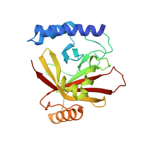
1NG5 | pdb_00001ng5

2.0 A crystal structure of Staphylococcus aureus Sortase B


Experimental Data Snapshot

  • Method: X-RAY DIFFRACTION
  • Resolution: 2.00 Å
  • R-Value Free: 
    0.248 (Depositor), 0.251 (DCC) 
  • R-Value Work: 
    0.237 (Depositor), 0.242 (DCC) 
  • R-Value Observed: 
    0.237 (Depositor) 

wwPDB Validation 3D Report Full Report

Validation slider image for 1NG5

This is version 1.5 of the entry. See complete history. 

Literature

Structures of sortase B from Staphylococcus aureus and Bacillus anthracis reveal catalytic amino acid triad in the active site.

Zhang, R., Wu, R., Joachimiak, G., Mazmanian, S.K., Missiakas, D.M., Gornicki, P., Schneewind, O., Joachimiak, A.

(2004) Structure 12: 1147-1156

  • DOI: https://doi.org/10.1016/j.str.2004.06.001
  • Primary Citation Related Structures: 
    1NG5, 1RZ2

  • PubMed Abstract: 

    Surface proteins attached by sortases to the cell wall envelope of bacterial pathogens play important roles during infection. Sorting and attachment of these proteins is directed by C-terminal signals. Sortase B of S. aureus recognizes a motif NPQTN, cleaves the polypeptide after the Thr residue, and attaches the protein to pentaglycine cross-bridges. Sortase B of B. anthracis is thought to recognize the NPKTG motif, and attaches surface proteins to m-diaminopimelic acid cross-bridges. We have determined crystal structure of sortase B from B. anthracis and S. aureus at 1.6 and 2.0 A resolutions, respectively. These structures show a beta-barrel fold with alpha-helical elements on its outside, a structure thus far exclusive to the sortase family. A putative active site located on the edge of the beta-barrel is comprised of a Cys-His-Asp catalytic triad and presumably faces the bacterial cell surface. A putative binding site for the sorting signal is located nearby.


  • Organizational Affiliation: 
    • Structural Biology Center and Midwest Center for Structural Genomics, Argonne National Laboratory, 9700 South Cass Avenue, Building 202, Argonne, IL 60439 USA.

Macromolecule Content 

  • Total Structure Weight: 51.28 kDa 
  • Atom Count: 3,799 
  • Modeled Residue Count: 417 
  • Deposited Residue Count: 430 
  • Unique protein chains: 1

Macromolecules

Find similar proteins by:|  3D Structure
Entity ID: 1
MoleculeChains  Sequence LengthOrganismDetailsImage
sortase B
A, B
215Staphylococcus aureus subsp. aureus N315Mutation(s): 0 
Gene Names: SA0982
EC: 3.4.22.71
UniProt
Find proteins for Q2FZE3 (Staphylococcus aureus (strain NCTC 8325 / PS 47))
Explore Q2FZE3 
Go to UniProtKB:  Q2FZE3
Entity Groups
Sequence Clusters30% Identity50% Identity70% Identity90% Identity95% Identity100% Identity
UniProt GroupQ2FZE3
Sequence Annotations
Expand
Reference Sequence

Experimental Data & Validation

Experimental Data

  • Method: X-RAY DIFFRACTION
  • Resolution: 2.00 Å
  • R-Value Free:  0.248 (Depositor), 0.251 (DCC) 
  • R-Value Work:  0.237 (Depositor), 0.242 (DCC) 
  • R-Value Observed: 0.237 (Depositor) 
Space Group: P 21 21 2
Unit Cell:
Length ( Å )Angle ( ˚ )
a = 71.158α = 90
b = 104.383β = 90
c = 58.158γ = 90
Software Package:
Software NamePurpose
CNSrefinement
d*TREKdata reduction
HKL-2000data scaling
CNSphasing

Structure Validation

View Full Validation Report


Entry History 

Deposition Data

Revision History  (Full details and data files)

  • Version 1.0: 2003-09-23
    Type: Initial release
  • Version 1.1: 2008-04-29
    Changes: Version format compliance
  • Version 1.2: 2011-07-13
    Changes: Source and taxonomy, Version format compliance
  • Version 1.3: 2017-10-11
    Changes: Refinement description
  • Version 1.4: 2022-12-21
    Changes: Database references
  • Version 1.5: 2024-05-22
    Changes: Data collection