1NF2 | pdb_00001nf2

X-ray crystal structure of TM0651 from Thermotoga maritima


Experimental Data Snapshot

  • Method: X-RAY DIFFRACTION
  • Resolution: 2.20 Å
  • R-Value Free: 
    0.263 (Depositor), 0.260 (DCC) 
  • R-Value Work: 
    0.217 (Depositor), 0.216 (DCC) 
  • R-Value Observed: 
    0.217 (Depositor) 

wwPDB Validation 3D Report Full Report

Validation slider image for 1NF2

This is version 1.3 of the entry. See complete history

Literature

Crystal structure of a phosphatase with a unique substrate binding domain from Thermotoga maritima

Shin, D.H.Roberts, A.Jancarik, J.Yokota, H.Kim, R.Wemmer, D.E.Kim, S.H.

(2003) Protein Sci 12: 1464-1472

  • DOI: https://doi.org/10.1110/ps.0302703
  • Primary Citation Related Structures: 
    1NF2

  • PubMed Abstract: 

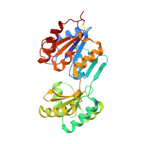
    We have determined the crystal structure of a phosphatase with a unique substrate binding domain from Thermotoga maritima, TM0651 (gi 4981173), at 2.2 A resolution by selenomethionine single-wavelength anomalous diffraction (SAD) techniques. TM0651 is a member of the haloacid dehalogenase (HAD) superfamily, with sequence homology to trehalose-6-phosphate phosphatase and sucrose-6(F)-phosphate phosphohydrolase. Selenomethionine labeled TM0651 crystallized in space group C2 with three monomers per asymmetric unit. Each monomer has approximate dimensions of 65 x 40 x 35 A(3), and contains two domains: a domain of known hydrolase fold characteristic of the HAD family, and a domain with a new tertiary fold consisting of a six-stranded beta-sheet surrounded by four alpha-helices. There is one disulfide bond between residues Cys35 and Cys265 in each monomer. One magnesium ion and one sulfate ion are bound in the active site. The superposition of active site residues with other HAD family members indicates that TM0651 is very likely a phosphatase that acts through the formation of a phosphoaspartate intermediate, which is supported by both NMR titration data and a biochemical assay. Structural and functional database searches and the presence of many aromatic residues in the interface of the two domains suggest the substrate of TM0651 is a carbohydrate molecule. From the crystal structure and NMR data, the protein likely undergoes a conformational change upon substrate binding.


  • Organizational Affiliation
    • Physical Biosciences Division, Lawrence Berkeley National Laboratory, Berkeley, CA 94720, USA.

Macromolecule Content 

  • Total Structure Weight: 94.33 kDa 
  • Atom Count: 7,085 
  • Modeled Residue Count: 801 
  • Deposited Residue Count: 804 
  • Unique protein chains: 1

Macromolecules

Find similar proteins by:|  3D Structure
Entity ID: 1
MoleculeChains  Sequence LengthOrganismDetailsImage
phosphatase
A, B, C
268Thermotoga maritimaMutation(s): 0 
UniProt
Find proteins for Q9WZB9 (Thermotoga maritima (strain ATCC 43589 / DSM 3109 / JCM 10099 / NBRC 100826 / MSB8))
Explore Q9WZB9 
Go to UniProtKB:  Q9WZB9
Entity Groups
Sequence Clusters30% Identity50% Identity70% Identity90% Identity95% Identity100% Identity
UniProt GroupQ9WZB9
Sequence Annotations
Expand
Reference Sequence

Small Molecules

Ligands 2 Unique
IDChains Name / Formula / InChI Key2D Diagram3D Interactions
SO4

Query on SO4


Download:Ideal Coordinates CCD File
D [auth A]
E [auth A]
G [auth B]
H [auth B]
J [auth C]
D [auth A],
E [auth A],
G [auth B],
H [auth B],
J [auth C],
K [auth C]
SULFATE ION
O4 S
QAOWNCQODCNURD-UHFFFAOYSA-L
MG

Query on MG


Download:Ideal Coordinates CCD File
F [auth A],
I [auth B],
L [auth C]
MAGNESIUM ION
Mg
JLVVSXFLKOJNIY-UHFFFAOYSA-N

Experimental Data & Validation

Experimental Data

  • Method: X-RAY DIFFRACTION
  • Resolution: 2.20 Å
  • R-Value Free:  0.263 (Depositor), 0.260 (DCC) 
  • R-Value Work:  0.217 (Depositor), 0.216 (DCC) 
  • R-Value Observed: 0.217 (Depositor) 
Space Group: C 1 2 1
Unit Cell:
Length ( Å )Angle ( ˚ )
a = 158.41α = 90
b = 93.88β = 127.91
c = 98.42γ = 90
Software Package:
Software NamePurpose
DENZOdata reduction
SCALEPACKdata scaling
SOLVEphasing
CNSrefinement

Structure Validation

View Full Validation Report


Entry History 

Deposition Data

Revision History  (Full details and data files)

  • Version 1.0: 2003-09-16
    Type: Initial release
  • Version 1.1: 2008-04-28
    Changes: Version format compliance
  • Version 1.2: 2011-07-13
    Changes: Derived calculations, Version format compliance
  • Version 1.3: 2024-11-13
    Changes: Data collection, Database references, Derived calculations, Structure summary