1NBE | pdb_00001nbe

ASPARTATE TRANSCARBAMOYLASE REGULATORY CHAIN MUTANT (T82A)


Experimental Data Snapshot

  • Method: X-RAY DIFFRACTION
  • Resolution: 2.60 Å
  • R-Value Free: 
    0.237 (Depositor) 
  • R-Value Work: 
    0.177 (Depositor) 
  • R-Value Observed: 
    0.177 (Depositor) 

Starting Model: experimental
View more details

wwPDB Validation 3D Report Full Report

Validation slider image for 1NBE

This is version 1.5 of the entry. See complete history

Literature

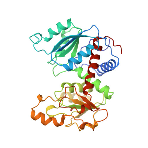
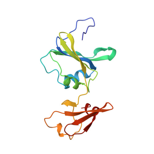
A single mutation in the regulatory chain of Escherichia coli aspartate transcarbamoylase results in an extreme T-state structure.

Williams, M.K.Stec, B.Kantrowitz, E.R.

(1998) J Mol Biology 281: 121-134

  • DOI: https://doi.org/10.1006/jmbi.1998.1923
  • Primary Citation Related Structures: 
    1NBE

  • PubMed Abstract: 

    Kinetic analysis of a mutant version of Escherichia coli aspartate transcarbamoylase in which Thr82 in the regulatory chain (Thr82r) was replaced by Ala results in a shift in the T <==> R equilibrium towards the T-state. In order to understand the structural determinants of this T-state stabilization, the X-ray structure of the unliganded Thr82r-->Ala enzyme was determined at 2. 6 A resolution and refined to a crystallographic residual of 0.175. The structure of the mutant r1 regulatory chain is more similar to that of the r6 regulatory chain than observed for the wild-type enzyme, resulting in a more symmetric structure. Furthermore, the structural changes in the mutant enzyme appears to occur only in the r1 chain, while the r6 chain is almost identical in structure to that of the r6 chain of the wild-type enzyme. The structure of the mutant enzyme exhibits alterations in the subunit interfaces between the regulatory and catalytic chains, as well as in the interface between the allosteric and zinc domains within the regulatory chain. Moreover, the regulatory dimers are rotated around their respective 2-fold axes approximately 1 degrees beyond the rotation which occurs in the wild-type T-state enzyme. The structural analysis indicates that the enzyme is an "extreme" T-state, in which a larger rotation of the regulatory dimers is required for the T to R transition compared to the wild-type enzyme. This extreme T-state structure correlates well with the kinetic parameters determined for the mutant enzyme, showing a stabilized T-state. Furthermore, the structural analysis of the mutant enzyme suggests that replacement of Thr82r with Ala alters the local conformation of the nucleotide binding pocket and therefore offers a plausible explanation for the reduced affinity of the enzyme for nucleotides.


  • Organizational Affiliation
    • Merkert Chemistry Center, Boston College, Chestnut Hill, MA 02167, USA.

Macromolecule Content 

  • Total Structure Weight: 103.57 kDa 
  • Atom Count: 7,816 
  • Modeled Residue Count: 926 
  • Deposited Residue Count: 926 
  • Unique protein chains: 2

Macromolecules

Find similar proteins by:|  3D Structure
Entity ID: 1
MoleculeChains  Sequence LengthOrganismDetailsImage
ASPARTATE TRANSCARBAMOYLASE
A, C
310Escherichia coliMutation(s): 0 
Gene Names: PURBI
EC: 2.1.3.2
UniProt
Find proteins for P0A786 (Escherichia coli (strain K12))
Explore P0A786 
Go to UniProtKB:  P0A786
Entity Groups
Sequence Clusters30% Identity50% Identity70% Identity90% Identity95% Identity100% Identity
UniProt GroupP0A786
Sequence Annotations
Expand
Reference Sequence
Find similar proteins by:|  3D Structure
Entity ID: 2
MoleculeChains  Sequence LengthOrganismDetailsImage
ASPARTATE TRANSCARBAMOYLASE
B, D
153Escherichia coliMutation(s): 1 
Gene Names: PURBI
EC: 2.1.3.2
UniProt
Find proteins for P0A7F3 (Escherichia coli (strain K12))
Explore P0A7F3 
Go to UniProtKB:  P0A7F3
Entity Groups
Sequence Clusters30% Identity50% Identity70% Identity90% Identity95% Identity100% Identity
UniProt GroupP0A7F3
Sequence Annotations
Expand
Reference Sequence

Experimental Data & Validation

Experimental Data

  • Method: X-RAY DIFFRACTION
  • Resolution: 2.60 Å
  • R-Value Free:  0.237 (Depositor) 
  • R-Value Work:  0.177 (Depositor) 
  • R-Value Observed: 0.177 (Depositor) 
Space Group: P 3 2 1
Unit Cell:
Length ( Å )Angle ( ˚ )
a = 122.47α = 90
b = 122.47β = 90
c = 142.77γ = 120
Software Package:
Software NamePurpose
X-PLORmodel building
X-PLORrefinement
SDMSdata reduction
SDMSdata scaling
X-PLORphasing

Structure Validation

View Full Validation Report


Entry History 

Deposition Data

Revision History  (Full details and data files)

  • Version 1.0: 1998-10-14
    Type: Initial release
  • Version 1.1: 2008-03-03
    Changes: Version format compliance
  • Version 1.2: 2011-07-13
    Changes: Derived calculations, Version format compliance
  • Version 1.3: 2021-11-03
    Changes: Database references, Derived calculations, Other
  • Version 1.4: 2023-08-09
    Changes: Refinement description
  • Version 1.5: 2024-05-22
    Changes: Data collection