1N51 | pdb_00001n51

Aminopeptidase P in complex with the inhibitor apstatin


Experimental Data Snapshot

  • Method: X-RAY DIFFRACTION
  • Resolution: 2.30 Å
  • R-Value Free: 
    0.204 (Depositor), 0.200 (DCC) 
  • R-Value Work: 
    0.179 (Depositor), 0.174 (DCC) 

Starting Model: experimental
View more details

wwPDB Validation 3D Report Full Report

Validation slider image for 1N51

This is version 2.0 of the entry. See complete history

Literature

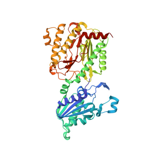
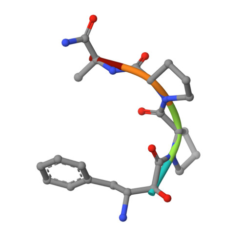
Structure of Escherichia coli aminopeptidase P in complex with the inhibitor apstatin.

Graham, S.C.Maher, M.J.Simmons, W.H.Freeman, H.C.Guss, J.M.

(2004) Acta Crystallogr D Biol Crystallogr 60: 1770-1779

  • DOI: https://doi.org/10.1107/S0907444904018724
  • Primary Citation Related Structures: 
    1N51

  • PubMed Abstract: 

    Aminopeptidase P (APPro) is a metalloprotease whose active site includes a dinuclear manganese(II) cluster. The enzyme cleaves the N-terminal residue from a polypeptide when the second residue is proline. A complex of Escherichia coli APPro (EcAPPro) with an inhibitor, apstatin [N-(2S,3R)-3-amino-2-hydroxy-4-phenyl-butanoyl-L-prolyl-L-prolyl-L-alaninamide], has been crystallized. Apstatin binds to the active site of EcAPPro with its N-terminal amino group coordinated to one of the two Mn(II) atoms at the metal centre. The apstatin hydroxyl group replaces a hydroxide ion which bridges the two metal atoms in the native enzyme. The first proline residue of apstatin lies in a small hydrophobic cleft. The structure of the apstatin-EcAPPro complex has been refined at 2.3 A resolution with residuals R = 0.179 and R(free) = 0.204. The structure of the complex illustrates how apstatin inhibits APPro and suggests how substrates may bind to the enzyme, but the basis of the proline-specificity remains elusive.


  • Organizational Affiliation
    • School of Molecular and Microbial Biosciences, University of Sydney, NSW 2006, Australia.

Macromolecule Content 

  • Total Structure Weight: 50.37 kDa 
  • Atom Count: 3,991 
  • Modeled Residue Count: 445 
  • Deposited Residue Count: 445 
  • Unique protein chains: 2

Macromolecules

Find similar proteins by:|  3D Structure
Entity ID: 1
MoleculeChains  Sequence LengthOrganismDetailsImage
Xaa-Pro aminopeptidase440Escherichia coliMutation(s): 0 
Gene Names: PEPP
EC: 3.4.11.9
UniProt
Find proteins for P15034 (Escherichia coli (strain K12))
Explore P15034 
Go to UniProtKB:  P15034
Entity Groups
Sequence Clusters30% Identity50% Identity70% Identity90% Identity95% Identity100% Identity
UniProt GroupP15034
Sequence Annotations
Expand
Reference Sequence
Find similar proteins by:  Sequence   |   3D Structure  
Entity ID: 2
MoleculeChains  Sequence LengthOrganismDetailsImage
apstatin5N/AMutation(s): 0 

Biologically Interesting Molecules (External Reference) 

1 Unique

Experimental Data & Validation

Experimental Data

  • Method: X-RAY DIFFRACTION
  • Resolution: 2.30 Å
  • R-Value Free:  0.204 (Depositor), 0.200 (DCC) 
  • R-Value Work:  0.179 (Depositor), 0.174 (DCC) 
Space Group: I 41 2 2
Unit Cell:
Length ( Å )Angle ( ˚ )
a = 139.319α = 90
b = 139.319β = 90
c = 231.005γ = 90
Software Package:
Software NamePurpose
CNSrefinement
DENZOdata reduction
SCALEPACKdata scaling
AMoREphasing

Structure Validation

View Full Validation Report


Entry History 

Deposition Data

Revision History  (Full details and data files)

  • Version 1.0: 2003-12-16
    Type: Initial release
  • Version 1.1: 2008-04-28
    Changes: Version format compliance
  • Version 1.2: 2011-07-13
    Changes: Atomic model, Database references, Derived calculations, Non-polymer description, Structure summary, Version format compliance
  • Version 1.3: 2012-12-12
    Changes: Other
  • Version 1.4: 2023-10-25
    Changes: Data collection, Database references, Derived calculations, Refinement description
  • Version 2.0: 2023-11-15
    Changes: Atomic model, Data collection, Derived calculations