1N3Y | pdb_00001n3y

Crystal structure of the alpha-X beta2 integrin I domain


Experimental Data Snapshot

  • Method: X-RAY DIFFRACTION
  • Resolution: 1.65 Å
  • R-Value Free: 
    0.225 (Depositor), 0.224 (DCC) 
  • R-Value Work: 
    0.208 (Depositor), 0.208 (DCC) 
  • R-Value Observed: 
    0.209 (Depositor) 

Starting Model: other
View more details

wwPDB Validation 3D Report Full Report

Validation slider image for 1N3Y

This is version 1.5 of the entry. See complete history

Literature

Structure and allosteric regulation of the alpha X beta 2 integrin I domain.

Vorup-Jensen, T.Ostermeier, C.Shimaoka, M.Hommel, U.Springer, T.A.

(2003) Proc Natl Acad Sci U S A 100: 1873-1878

  • DOI: https://doi.org/10.1073/pnas.0237387100
  • Primary Citation Related Structures: 
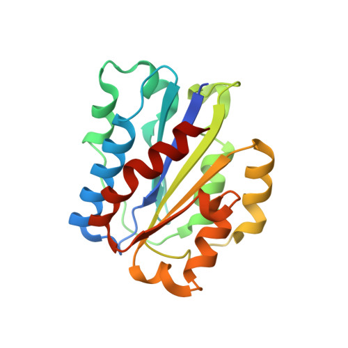
    1N3Y

  • PubMed Abstract: 

    The integrin alpha X beta 2 (CD11c/CD18, p150,95) binds ligands through the I domain of the alpha X subunit. Ligands include the complement factor fragment iC3b, a key component in the innate immune defense, which, together with the expression of alpha X beta 2 on dendritic cells and on other leukocytes, suggests a role in the immune response. We now report the structure of the alpha X I domain resolved at 1.65 A by x-ray crystallography. To analyze structural requirements for ligand binding we made a mutation in the alpha X I domain C-terminal helix, which increased the affinity for iC3b approximately 200-fold to 2.4 microM compared with the wild-type domain affinity of approximately 400 microM. Gel permeation chromatography supported a conformational change between the wild-type and mutated domains. Conservation of allosteric regulation in the alpha X I domain points to the functional importance of this phenomenon.


  • Organizational Affiliation
    • Center for Blood Research, Department of Pathology, Harvard Medical School, 200 Longwood Avenue, Boston, MA 02115, USA.

Macromolecule Content 

  • Total Structure Weight: 22.36 kDa 
  • Atom Count: 1,803 
  • Modeled Residue Count: 189 
  • Deposited Residue Count: 198 
  • Unique protein chains: 1

Macromolecules

Find similar proteins by:|  3D Structure
Entity ID: 1
MoleculeChains  Sequence LengthOrganismDetailsImage
Integrin alpha-X198Homo sapiensMutation(s): 2 
Gene Names: ITGAX OR CD11C
UniProt & NIH Common Fund Data Resources
Find proteins for P20702 (Homo sapiens)
Explore P20702 
Go to UniProtKB:  P20702
PHAROS:  P20702
GTEx:  ENSG00000140678 
Entity Groups
Sequence Clusters30% Identity50% Identity70% Identity90% Identity95% Identity100% Identity
UniProt GroupP20702
Sequence Annotations
Expand
Reference Sequence

Experimental Data & Validation

Experimental Data

  • Method: X-RAY DIFFRACTION
  • Resolution: 1.65 Å
  • R-Value Free:  0.225 (Depositor), 0.224 (DCC) 
  • R-Value Work:  0.208 (Depositor), 0.208 (DCC) 
  • R-Value Observed: 0.209 (Depositor) 
Space Group: P 61
Unit Cell:
Length ( Å )Angle ( ˚ )
a = 84.694α = 90
b = 84.694β = 90
c = 65.776γ = 120
Software Package:
Software NamePurpose
DENZOdata reduction
SCALEPACKdata scaling
CNSrefinement
CNXrefinement
CNSphasing

Structure Validation

View Full Validation Report


Entry History 

Deposition Data

Revision History  (Full details and data files)

  • Version 1.0: 2003-02-18
    Type: Initial release
  • Version 1.1: 2008-04-28
    Changes: Version format compliance
  • Version 1.2: 2011-07-13
    Changes: Version format compliance
  • Version 1.3: 2021-10-27
    Changes: Database references
  • Version 1.4: 2024-02-14
    Changes: Data collection
  • Version 1.5: 2024-04-03
    Changes: Refinement description