1N1H | pdb_00001n1h

Initiation complex of polymerase lambda3 from reovirus


Experimental Data Snapshot

  • Method: X-RAY DIFFRACTION
  • Resolution: 2.80 Å
  • R-Value Free: 
    0.268 (Depositor), 0.262 (DCC) 
  • R-Value Work: 
    0.216 (Depositor), 0.206 (DCC) 
  • R-Value Observed: 
    0.216 (Depositor) 

Starting Model: experimental
View more details

wwPDB Validation 3D Report Full Report

Validation slider image for 1N1H

Ligand Structure Quality Assessment 


This is version 1.4 of the entry. See complete history

Literature

RNA Synthesis in a Cage--Structural Studies of Reovirus Polymerase [lambda] 3

Tao, Y.Farsetta, D.L.Nibert, M.L.Harrison, S.C.

(2002) Cell 111: 733-745

  • DOI: https://doi.org/10.1016/s0092-8674(02)01110-8
  • Primary Citation Related Structures: 
    1MUK, 1MWH, 1N1H, 1N35, 1N38

  • PubMed Abstract: 

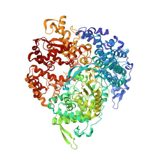
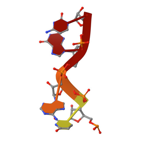
    The reovirus polymerase and those of other dsRNA viruses function within the confines of a protein capsid to transcribe the tightly packed dsRNA genome segments. The crystal structure of the reovirus polymerase, lambda3, determined at 2.5 A resolution, shows a fingers-palm-thumb core, similar to those of other viral polymerases, surrounded by major N- and C-terminal elaborations, which create a cage-like structure, with four channels leading to the catalytic site. This "caged" polymerase has allowed us to visualize the results of several rounds of RNA polymerization directly in the crystals. A 5' cap binding site on the surface of lambda3 suggests a template retention mechanism by which attachment of the 5' end of the plus-sense strand facilitates insertion of the 3' end of the minus-sense strand into the template channel.


  • Organizational Affiliation
    • Howard Hughes Medical Institute, Department of Molecular and Cellular Biology, Harvard University, Cambridge, MA 02138, USA.

Macromolecule Content 

  • Total Structure Weight: 146.21 kDa 
  • Atom Count: 10,367 
  • Modeled Residue Count: 1,268 
  • Deposited Residue Count: 1,273 
  • Unique protein chains: 1
  • Unique nucleic acid chains: 1

Macromolecules


Find similar proteins by:|  3D Structure
Entity ID: 2
MoleculeChains  Sequence LengthOrganismDetailsImage
Minor core protein lambda 3B [auth A]1,267Mammalian orthoreovirus 3 DearingMutation(s): 0 
Gene Names: L1
EC: 2.7.7.48
UniProt
Find proteins for P0CK31 (Reovirus type 3 (strain Dearing))
Explore P0CK31 
Go to UniProtKB:  P0CK31
Entity Groups
Sequence Clusters30% Identity50% Identity70% Identity90% Identity95% Identity100% Identity
UniProt GroupP0CK31
Sequence Annotations
Expand
Reference Sequence
Find similar nucleic acids by:  Sequence
Entity ID: 1
MoleculeChains LengthOrganismImage
5'-R(*AP*UP*UP*AP*GP*C)-3'A [auth B]6N/A
Sequence Annotations
Expand
Reference Sequence

Small Molecules

Ligands 5 Unique
IDChains Name / Formula / InChI Key2D Diagram3D Interactions
GH3

Query on GH3


Download:Ideal Coordinates CCD File
H [auth A]3'-DEOXY-GUANOSINE-5'-TRIPHOSPHATE
C10 H16 N5 O13 P3
QGYIFQKZZSSUCR-OBXARNEKSA-N
CH1

Query on CH1


Download:Ideal Coordinates CCD File
G [auth A]3'-DEOXY-CYTIDINE-5'-TRIPHOSPHATE
C9 H16 N3 O13 P3
CHKFLBOLYREYDO-SHYZEUOFSA-N
GDP

Query on GDP


Download:Ideal Coordinates CCD File
F [auth A]GUANOSINE-5'-DIPHOSPHATE
C10 H15 N5 O11 P2
QGWNDRXFNXRZMB-UUOKFMHZSA-N
G7M

Query on G7M


Download:Ideal Coordinates CCD File
E [auth A]N7-METHYL-GUANOSINE-5'-MONOPHOSPHATE
C11 H17 N5 O8 P
AOKQNZVJJXPUQA-KQYNXXCUSA-O
MN

Query on MN


Download:Ideal Coordinates CCD File
C [auth A],
D [auth A]
MANGANESE (II) ION
Mn
WAEMQWOKJMHJLA-UHFFFAOYSA-N

Experimental Data & Validation

Experimental Data

  • Method: X-RAY DIFFRACTION
  • Resolution: 2.80 Å
  • R-Value Free:  0.268 (Depositor), 0.262 (DCC) 
  • R-Value Work:  0.216 (Depositor), 0.206 (DCC) 
  • R-Value Observed: 0.216 (Depositor) 
Space Group: P 21 21 21
Unit Cell:
Length ( Å )Angle ( ˚ )
a = 70.883α = 90
b = 85.013β = 90
c = 249.294γ = 90
Software Package:
Software NamePurpose
DENZOdata reduction
SCALEPACKdata scaling
CNSrefinement
CNSphasing

Structure Validation

View Full Validation Report


Ligand Structure Quality Assessment 


Entry History 

Deposition Data

Revision History  (Full details and data files)

  • Version 1.0: 2002-12-25
    Type: Initial release
  • Version 1.1: 2008-04-28
    Changes: Version format compliance
  • Version 1.2: 2011-07-13
    Changes: Version format compliance
  • Version 1.3: 2024-02-14
    Changes: Data collection, Database references, Derived calculations
  • Version 1.4: 2024-04-03
    Changes: Refinement description