1MZS | pdb_00001mzs

CRYSTAL STRUCTURE OF BETA-KETOACYL-ACP SYNTHASE III WITH BOUND dichlorobenzyloxy-indole-carboxylic acid inhibitor


Experimental Data Snapshot

  • Method: X-RAY DIFFRACTION
  • Resolution: 2.10 Å
  • R-Value Free: 
    0.230 (Depositor) 
  • R-Value Work: 
    0.175 (Depositor) 
  • R-Value Observed: 
    0.175 (Depositor) 

Starting Model: experimental
View more details

wwPDB Validation 3D Report Full Report

Validation slider image for 1MZS

This is version 1.4 of the entry. See complete history. 

Literature

First X-ray cocrystal structure of a bacterial FabH condensing enzyme and a small molecule inhibitor achieved using rational design and homology modeling

Daines, R.A., Pendrak, I., Sham, K., Van Aller, G.S., Konstantinidis, A.K., Lonsdale, J.T., Janson, C.A., Qiu, X., Brandt, M., Khandekar, S.S., Silverman, C., Head, M.S.

(2003) J Med Chem 46: 5-8

  • DOI: https://doi.org/10.1021/jm025571b
  • Primary Citation Related Structures: 
    1MZS

  • PubMed Abstract: 

    The first cocrystal structure of a bacterial FabH condensing enzyme and a small molecule inhibitor is reported. The inhibitor was obtained by rational modification of a high throughput screening lead with the aid of a S. pneumoniae FabH homology model. This homology model was used to design analogues that would have both high affinity for the enzyme and appropriate aqueous solubility to facilitate cocrystallization studies.


  • Organizational Affiliation: 
    • Department of Medicinal Chemistry, GlaxoSmithKline, 1250 S. Collegeville Road, Collegeville, Pennsylvania 19426, USA.

Macromolecule Content 

  • Total Structure Weight: 34.14 kDa 
  • Atom Count: 2,559 
  • Modeled Residue Count: 317 
  • Deposited Residue Count: 317 
  • Unique protein chains: 1

Macromolecules

Find similar proteins by:|  3D Structure
Entity ID: 1
MoleculeChains  Sequence LengthOrganismDetailsImage
3-oxoacyl-[acyl-carrier-protein] synthase III317Escherichia coliMutation(s): 1 
EC: 2.3.1.41 (PDB Primary Data), 2.3.1.180 (UniProt)
UniProt
Find proteins for P0A6R0 (Escherichia coli (strain K12))
Explore P0A6R0 
Go to UniProtKB:  P0A6R0
Entity Groups
Sequence Clusters30% Identity50% Identity70% Identity90% Identity95% Identity100% Identity
UniProt GroupP0A6R0
Sequence Annotations
Expand
Reference Sequence

Small Molecules

Ligands 2 Unique
IDChains Name / Formula / InChI Key2D Diagram3D Interactions
669

Query on 669


Download:Ideal Coordinates CCD File
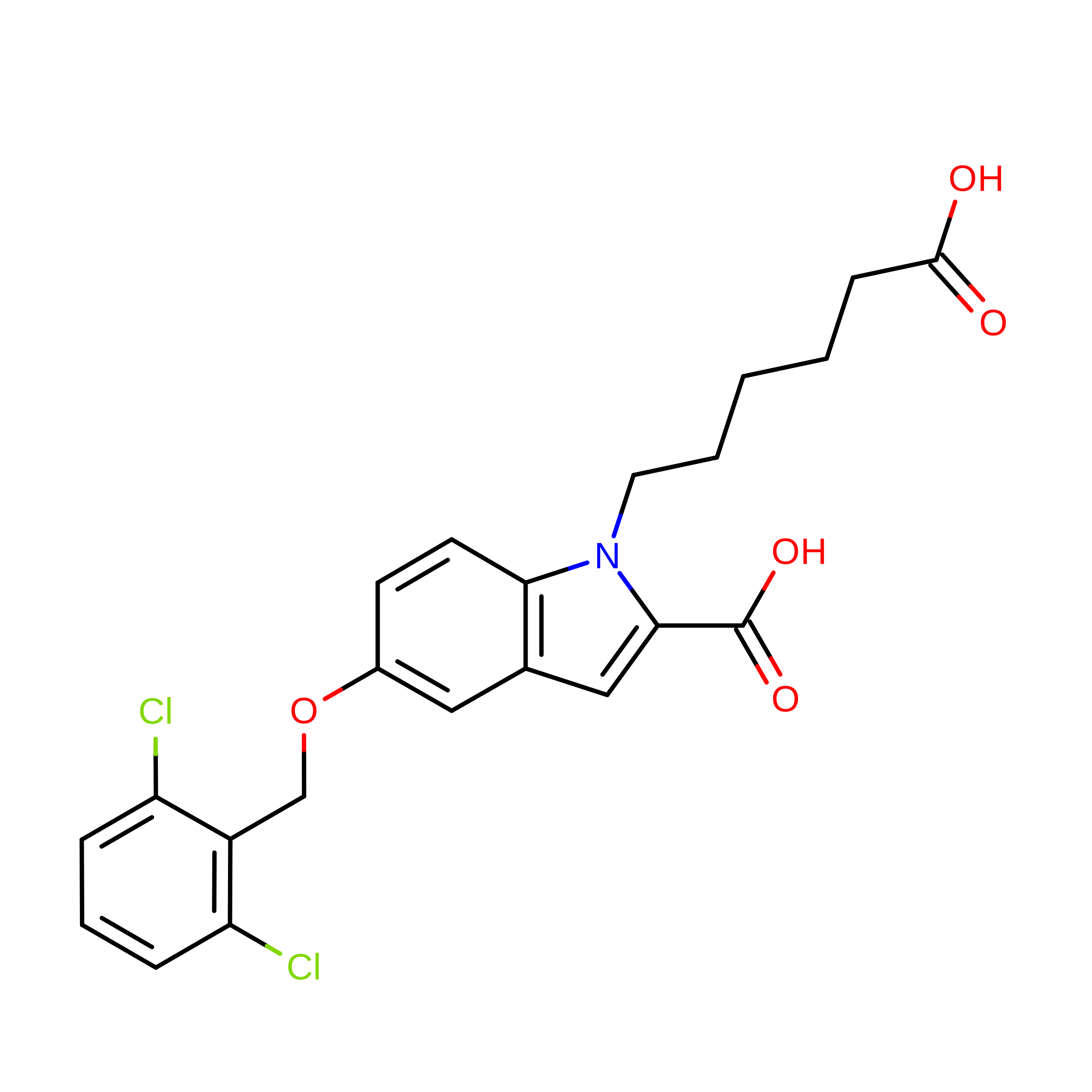
C [auth A]1-(5-CARBOXYPENTYL)-5-(2,6-DICHLOROBENZYLOXY)-1H-INDOLE-2-CARBOXYLIC ACID
C22 H21 Cl2 N O5
JTGPYFFQVOIJKR-UHFFFAOYSA-N
PO4

Query on PO4


Download:Ideal Coordinates CCD File
B [auth A]PHOSPHATE ION
O4 P
NBIIXXVUZAFLBC-UHFFFAOYSA-K
Modified Residues  1 Unique
IDChains TypeFormula2D DiagramParent
OCS
Query on OCS
A
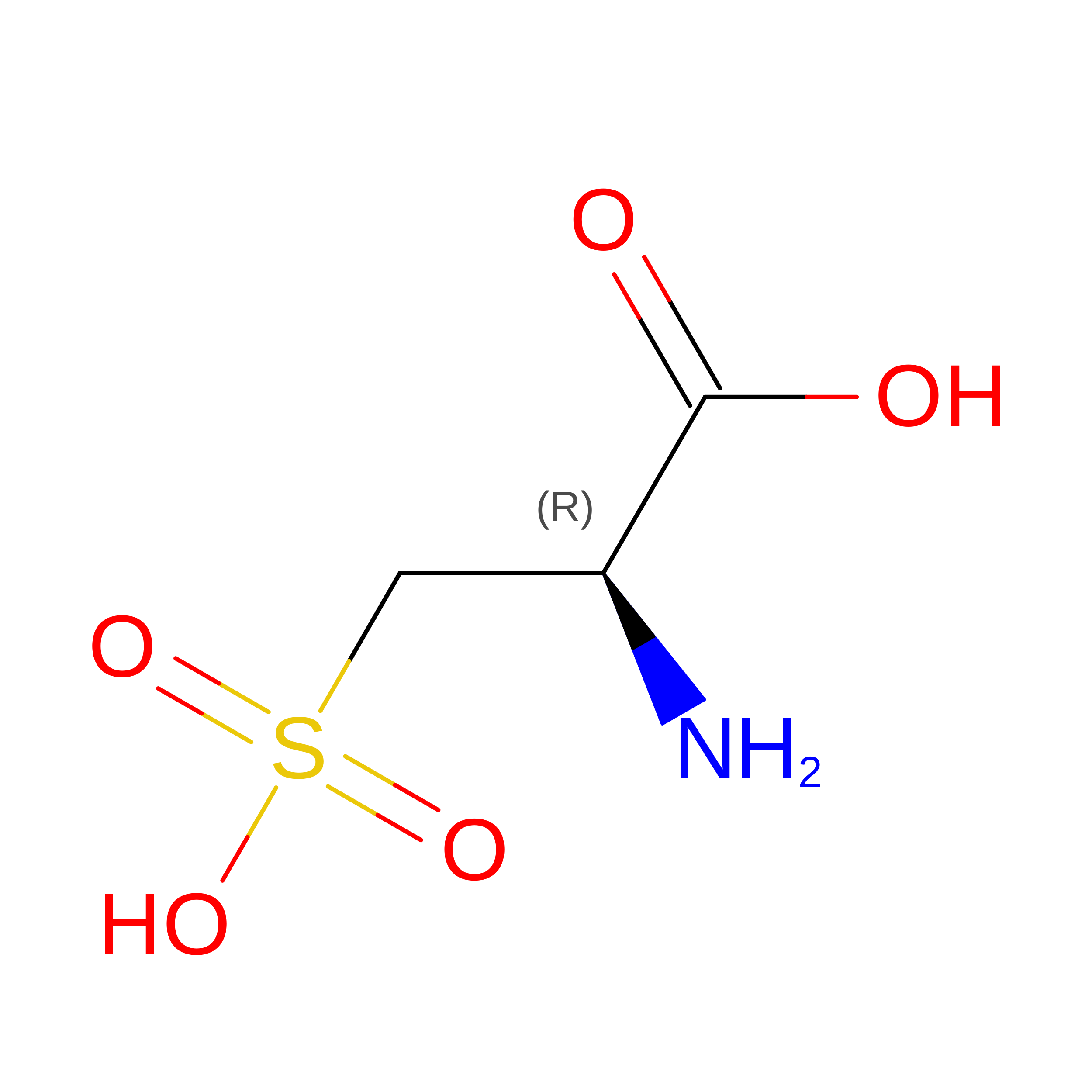
L-PEPTIDE LINKINGC3 H7 N O5 SCYS

Experimental Data & Validation

Experimental Data

  • Method: X-RAY DIFFRACTION
  • Resolution: 2.10 Å
  • R-Value Free:  0.230 (Depositor) 
  • R-Value Work:  0.175 (Depositor) 
  • R-Value Observed: 0.175 (Depositor) 
Space Group: P 41 21 2
Unit Cell:
Length ( Å )Angle ( ˚ )
a = 72.34α = 90
b = 72.34β = 90
c = 100.73γ = 90
Software Package:
Software NamePurpose
MAR345data collection
SCALEPACKdata scaling
X-PLORmodel building
CNSrefinement
X-PLORphasing

Structure Validation

View Full Validation Report


Entry History 

Revision History  (Full details and data files)

  • Version 1.0: 2002-11-13
    Type: Initial release
  • Version 1.1: 2008-04-28
    Changes: Version format compliance
  • Version 1.2: 2011-07-13
    Changes: Derived calculations, Version format compliance
  • Version 1.3: 2017-10-11
    Changes: Refinement description
  • Version 1.4: 2025-03-26
    Changes: Data collection, Database references, Derived calculations, Refinement description, Structure summary