1MZB | pdb_00001mzb

Ferric uptake regulator


Experimental Data Snapshot

  • Method: X-RAY DIFFRACTION
  • Resolution: 1.80 Å
  • R-Value Free: 
    0.254 (Depositor), 0.255 (DCC) 
  • R-Value Work: 
    0.226 (Depositor), 0.226 (DCC) 

wwPDB Validation 3D Report Full Report

Validation slider image for 1MZB

This is version 1.4 of the entry. See complete history

Literature

Architecture of a protein central to iron homeostatis: Crystal structure and spectroscopic analysis of the Ferric uptake regulator

Pohl, E.Haller, J.C.Mijovilovich, A.Meyer-Klaucke, W.Garman, E.Vasil, M.L.

(2003) Mol Microbiol 47: 903-915

  • DOI: https://doi.org/10.1046/j.1365-2958.2003.03337.x
  • Primary Citation Related Structures: 
    1MZB

  • PubMed Abstract: 

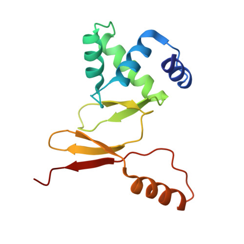
    Iron is an essential element for almost all organisms, although an overload of this element results in toxicity because of the formation of hydroxyl radicals. Consequently, most living entities have developed sophisticated mechanisms to control their intracellular iron concentration. In many bacteria, including the opportunistic pathogen Pseudomonas aeruginosa, this task is performed by the ferric uptake regulator (Fur). Fur controls a wide variety of basic physiological processes including iron uptake systems and the expression of exotoxin A. Here, we present the first crystal structure of Fur from P. aeruginosa in complex with Zn2+ determined at a resolution of 1.8 A. Furthermore, X-ray absorption spectroscopic measurements and microPIXE analysis were performed in order to characterize the distinct zinc and iron binding sites in solution. The combination of these complementary techniques enables us to present a model for the activation and DNA binding of the Fur protein.


  • Organizational Affiliation
    • European Molecular Biology Laboratory, Hamburg Outstation, Notkestr. 85, D-22603 Hamburg, Germany.

Macromolecule Content 

  • Total Structure Weight: 15.67 kDa 
  • Atom Count: 1,168 
  • Modeled Residue Count: 133 
  • Deposited Residue Count: 136 
  • Unique protein chains: 1

Macromolecules

Find similar proteins by:|  3D Structure
Entity ID: 1
MoleculeChains  Sequence LengthOrganismDetailsImage
ferric uptake regulation protein136Pseudomonas aeruginosaMutation(s): 0 
UniProt
Find proteins for Q03456 (Pseudomonas aeruginosa (strain ATCC 15692 / DSM 22644 / CIP 104116 / JCM 14847 / LMG 12228 / 1C / PRS 101 / PAO1))
Explore Q03456 
Go to UniProtKB:  Q03456
Entity Groups
Sequence Clusters30% Identity50% Identity70% Identity90% Identity95% Identity100% Identity
UniProt GroupQ03456
Sequence Annotations
Expand
Reference Sequence

Experimental Data & Validation

Experimental Data

  • Method: X-RAY DIFFRACTION
  • Resolution: 1.80 Å
  • R-Value Free:  0.254 (Depositor), 0.255 (DCC) 
  • R-Value Work:  0.226 (Depositor), 0.226 (DCC) 
Space Group: I 41 2 2
Unit Cell:
Length ( Å )Angle ( ˚ )
a = 63.3α = 90
b = 63.3β = 90
c = 180.1γ = 90
Software Package:
Software NamePurpose
MAR345data collection
DENZOdata reduction
SHELXSphasing
CNSrefinement
SCALEPACKdata scaling

Structure Validation

View Full Validation Report


Entry History 

Deposition Data

Revision History  (Full details and data files)

  • Version 1.0: 2003-10-07
    Type: Initial release
  • Version 1.1: 2008-04-28
    Changes: Version format compliance
  • Version 1.2: 2011-07-13
    Changes: Derived calculations, Version format compliance
  • Version 1.3: 2017-10-11
    Changes: Refinement description
  • Version 1.4: 2024-02-14
    Changes: Data collection, Database references, Derived calculations