1MW4 | pdb_00001mw4

Solution structure of the human Grb7-SH2 domain in complex with a 10 amino acid peptide pY1139


Experimental Data Snapshot

  • Method: SOLUTION NMR
  • Conformers Calculated: 50 
  • Conformers Submitted: 10 
  • Selection Criteria: structures with favorable non-bond energy 

wwPDB Validation 3D Report Full Report

Validation slider image for 1MW4

This is version 1.4 of the entry. See complete history

Literature

Solution structure of the human Grb7-SH2 domain/erbB2 peptide complex and structural basis for Grb7 binding to ErbB2

Ivancic, M.Daly, R.J.Lyons, B.A.

(2003) J Biomol NMR 27: 205-219

  • DOI: https://doi.org/10.1023/a:1025498409113
  • Primary Citation Related Structures: 
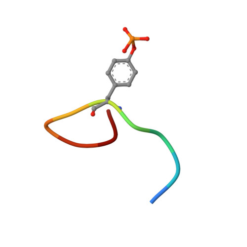
    1MW4

  • PubMed Abstract: 

    The solution structure of the hGrb7-SH2 domain in complex with a ten amino acid phosphorylated peptide ligand representative of the erbB2 receptor tyrosine kinase (pY1139) is presented as determined by nuclear magnetic resonance methods. The hGrb7-SH2 domain structure reveals the Src homology 2 domain topology consisting of a central beta-sheet capped at each end by an alpha-helix. The presence of a four residue insertion in the region between beta-strand E and the EF loop and resulting influences on the SH2 domain/peptide complex structure are discussed. The binding conformation of the erbB2 peptide is in a beta-turn similar to that found in phosphorylated tyrosine peptides bound to the Grb2-SH2 domain. To our knowledge this is only the second example of an SH2 domain binding its naturally occurring ligands in a turn, instead of extended, conformation. Close contacts between residues responsible for binding specificity in hGrb7-SH2 and the erbB2 peptide are characterized and the potential effect of mutation of these residues on the hGrb7-SH2 domain structure is discussed.


  • Organizational Affiliation
    • Department of Biochemistry, University of Vermont College of Medicine Burlington, VT 05405, U.S.A.

Macromolecule Content 

  • Total Structure Weight: 14.96 kDa 
  • Atom Count: 1,048 
  • Modeled Residue Count: 130 
  • Deposited Residue Count: 130 
  • Unique protein chains: 2

Macromolecules

Find similar proteins by:|  3D Structure
Entity ID: 1
MoleculeChains  Sequence LengthOrganismDetailsImage
Growth factor receptor-bound protein 7120Homo sapiensMutation(s): 0 
UniProt & NIH Common Fund Data Resources
Find proteins for Q14451 (Homo sapiens)
Explore Q14451 
Go to UniProtKB:  Q14451
PHAROS:  Q14451
GTEx:  ENSG00000141738 
Entity Groups
Sequence Clusters30% Identity50% Identity70% Identity90% Identity95% Identity100% Identity
UniProt GroupQ14451
Sequence Annotations
Expand
Reference Sequence
Find similar proteins by:  Sequence   |   3D Structure  
Entity ID: 2
MoleculeChains  Sequence LengthOrganismDetailsImage
Receptor protein-tyrosine kinase erbB-210N/AMutation(s): 1 
EC: 2.7.10.1
UniProt & NIH Common Fund Data Resources
Find proteins for P04626 (Homo sapiens)
Explore P04626 
Go to UniProtKB:  P04626
PHAROS:  P04626
GTEx:  ENSG00000141736 
Entity Groups
Sequence Clusters30% Identity50% Identity70% Identity90% Identity95% Identity100% Identity
UniProt GroupP04626
Sequence Annotations
Expand
Reference Sequence

Small Molecules

Modified Residues  1 Unique
IDChains TypeFormula2D DiagramParent
PTR
Query on PTR
B
L-PEPTIDE LINKINGC9 H12 N O6 PTYR

Experimental Data & Validation

Experimental Data

  • Method: SOLUTION NMR
  • Conformers Calculated: 50 
  • Conformers Submitted: 10 
  • Selection Criteria: structures with favorable non-bond energy 

Structure Validation

View Full Validation Report


Entry History 

Deposition Data

Revision History  (Full details and data files)

  • Version 1.0: 2003-09-09
    Type: Initial release
  • Version 1.1: 2008-04-28
    Changes: Version format compliance
  • Version 1.2: 2011-07-13
    Changes: Version format compliance
  • Version 1.3: 2012-05-02
    Changes: Structure summary
  • Version 1.4: 2024-10-16
    Changes: Data collection, Database references, Derived calculations, Structure summary