1MVM | pdb_00001mvm

MVM(STRAIN I), COMPLEX(VIRAL COAT/DNA), VP2, PH=7.5, T=4 DEGREES C


Experimental Data Snapshot

  • Method: X-RAY DIFFRACTION
  • Resolution: 3.50 Å

Starting Model: experimental
View more details

wwPDB Validation 3D Report Full Report

Validation slider image for 1MVM

This is version 2.1 of the entry. See complete history

Literature

Structure determination of minute virus of mice.

Llamas-Saiz, A.L.Agbandje-McKenna, M.Wikoff, W.R.Bratton, J.Tattersall, P.Rossmann, M.G.

(1997) Acta Crystallogr D Biol Crystallogr 53: 93-102

  • DOI: https://doi.org/10.1107/S0907444996010566
  • Primary Citation Related Structures: 
    1MVM

  • PubMed Abstract: 

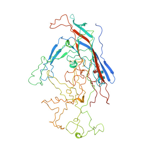
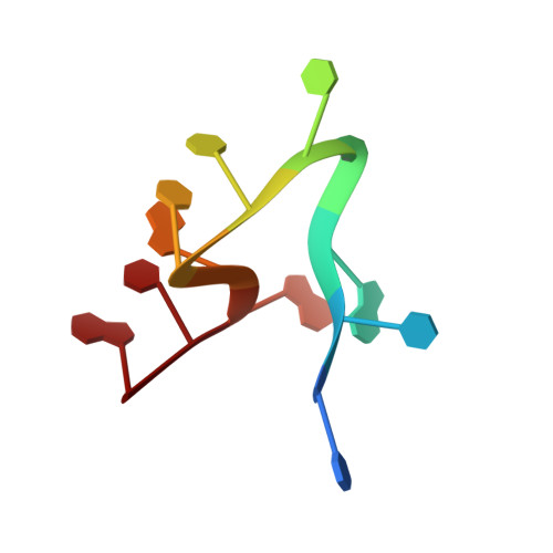
    The three-dimensional crystal structure of the single-stranded DNA-containing ('full') parvovirus, minute virus of mice (MVM), has been determined to 3.5 A resolution. Both full and empty particles of MVM were crystallized in the monoclinic space group C2 with cell dimensions of a = 448.7, b = 416.7, c = 305.3 A and beta = 95.8 degrees. Diffraction data were collected at the Cornell High Energy Synchrotron Source using an oscillation camera. The crystals have a pseudo higher R32 space group in which the particles are situated at two special positions with 32 point symmetry, separated by (1/2)c in the hexagonal setting. The self-rotation function showed that the particles are rotated with respect to each other by 60 degrees around the pseudo threefold axis. Subsequently, a more detailed analysis of the structure amplitudes demonstrated that the correct space-group symmetry is C2 as given above. Only one of the three twofold axes perpendicular to the threefold axis in the pseudo R32 space group is a 'true' crystallographic twofold axis; the other two are only 'local' non-crystallographic symmetry axes. The known canine parvovirus (CPV) structure was used as a phasing model to initiate real-space electron-density averaging phase improvement. The electron density was easily interpretable and clearly showed the amino-acid differences between MVM and CPV, although the final overall correlation coefficient was only 0.63. The structure of MVM has a large amount of icosahedrally ordered DNA, amounting to 22% of the viral genome, which is significantly more than that found in CPV.


  • Organizational Affiliation
    • Department of Biological Sciences, Purdue University, West Lafayette, IN 47907-1392, USA.

Macromolecule Content 

  • Total Structure Weight: 69.49 kDa 
  • Atom Count: 4,649 
  • Modeled Residue Count: 565 
  • Deposited Residue Count: 603 
  • Unique protein chains: 1
  • Unique nucleic acid chains: 3

Macromolecules


Find similar proteins by:|  3D Structure
Entity ID: 1
MoleculeChains  Sequence LengthOrganismDetailsImage
PROTEIN (MURINE MINUTE VIRUS COAT PROTEIN)587Minute virus of miceMutation(s): 2 
UniProt
Find proteins for P07302 (Murine minute virus (strain MVMi))
Explore P07302 
Go to UniProtKB:  P07302
Entity Groups
Sequence Clusters30% Identity50% Identity70% Identity90% Identity95% Identity100% Identity
UniProt GroupP07302
Sequence Annotations
Expand
Reference Sequence
Find similar nucleic acids by:  Sequence
Entity ID: 2
MoleculeChains LengthOrganismImage
DNA (5'-D(*CP*CP*AP*CP*CP*CP*CP*AP*AP*CP*A)-3')11N/A
Sequence Annotations
Expand
Reference Sequence
Find similar nucleic acids by:  Sequence
Entity ID: 3
MoleculeChains LengthOrganismImage
DNA (5'-D(*CP*AP*AP*A)-3')4N/A
Sequence Annotations
Expand
Reference Sequence
Find similar nucleic acids by:  Sequence
Entity ID: 4
MoleculeChains LengthOrganismImage
DNA (5'-D(P*A)-3')1N/A
Sequence Annotations
Expand
Reference Sequence

Experimental Data & Validation

Experimental Data

  • Method: X-RAY DIFFRACTION
  • Resolution: 3.50 Å
Space Group: C 1 2 1
Unit Cell:
Length ( Å )Angle ( ˚ )
a = 448.7α = 90
b = 416.7β = 95.8
c = 305.3γ = 90
Software Package:
Software NamePurpose
PURDUEmodel building
NONErefinement
PURDUEdata reduction
PURDUEdata scaling
PURDUEphasing

Structure Validation

View Full Validation Report


Entry History 

Deposition Data

Revision History  (Full details and data files)

  • Version 1.0: 1998-02-25
    Type: Initial release
  • Version 1.1: 2008-05-22
    Changes: Version format compliance
  • Version 1.2: 2011-07-13
    Changes: Version format compliance
  • Version 2.0: 2023-04-19
    Type: Remediation
    Changes: Advisory, Atomic model, Data collection, Database references, Derived calculations, Other, Refinement description
  • Version 2.1: 2024-04-03
    Changes: Data collection, Refinement description