1MUI | pdb_00001mui

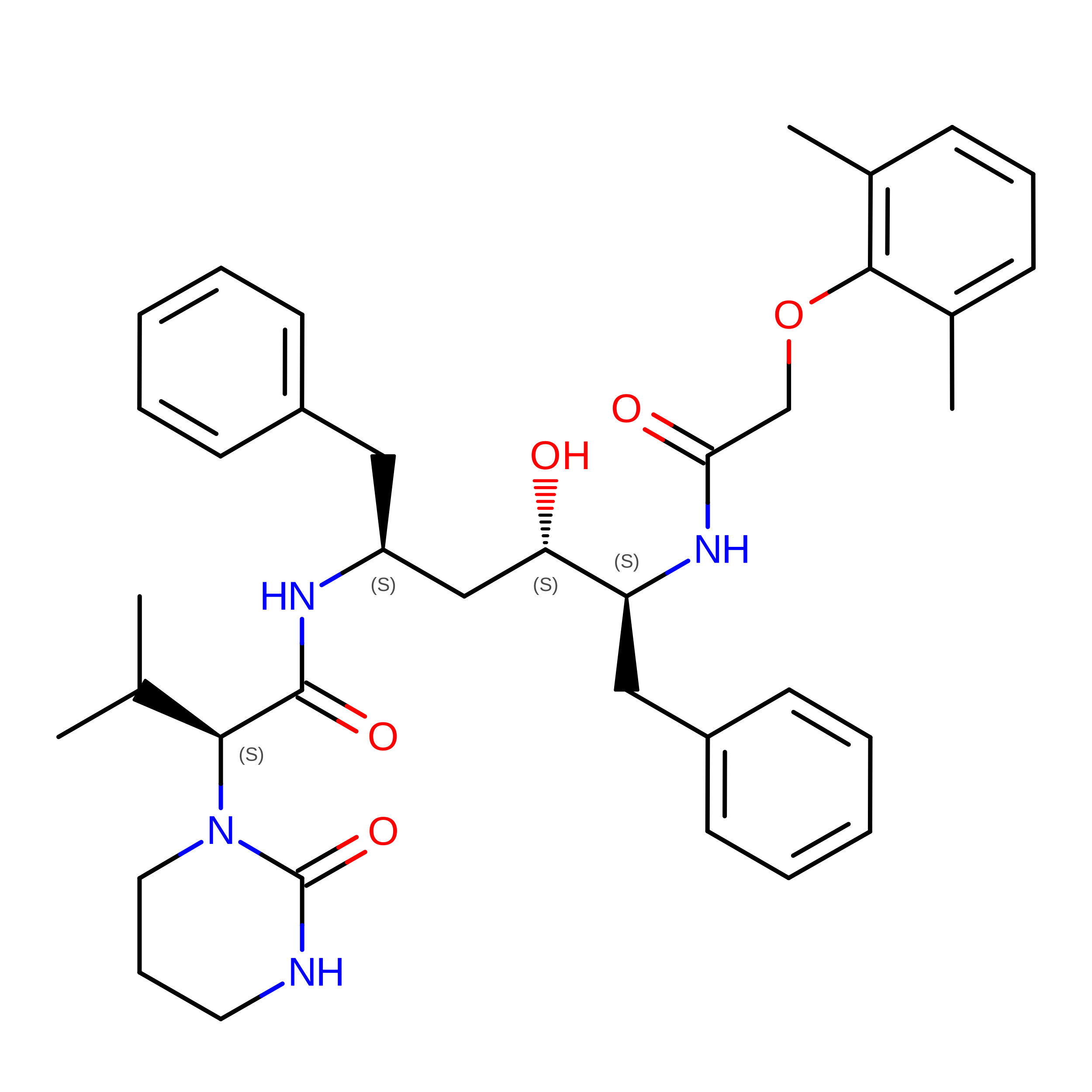
Crystal structure of HIV-1 protease complexed with Lopinavir.


Experimental Data Snapshot

  • Method: X-RAY DIFFRACTION
  • Resolution: 2.80 Å
  • R-Value Free: 
    0.329 (Depositor), 0.185 (DCC) 
  • R-Value Work: 
    0.261 (Depositor), 0.200 (DCC) 
  • R-Value Observed: 
    0.309 (Depositor) 

Starting Model: experimental
View more details

wwPDB Validation 3D Report Full Report

Validation slider image for 1MUI

Ligand Structure Quality Assessment 


This is version 1.4 of the entry. See complete history. 

Literature

X-ray Crystallographic Structure of ABT-378 (Lopinavir) Bound to HIV-1 Protease

Stoll, V., Qin, W., Stewart, K.D., Jakob, C., Park, C., Walter, K., Simmer, R.L., Helfrich, R., Bussiere, D., Kao, J., Kempf, D., Sham, H.L., Norbeck, D.W.

(2002) Bioorg Med Chem 10: 2803-2806

  • DOI: https://doi.org/10.1016/s0968-0896(02)00051-2
  • Primary Citation Related Structures: 
    1MUI

  • PubMed Abstract: 

    The crystal structure of ABT-378 (lopinavir), bound to the active site of HIV-1 protease is described. A comparison with crystal structures of ritonavir, A-78791, and BILA-2450 shows some analogous features with previous reported compounds. A cyclic urea unit in the P(2) position of ABT-378 is novel and makes two bidentate hydrogen bonds with Asp 29 of HIV-1 protease. In addition, a previously unreported shift in the Gly 48 carbonyl position is observed. A discussion of the structural features responsible for its high potency against wild-type HIV protease is given along with an analysis of the effect of active site mutations on potency in in vitro assays.


  • Organizational Affiliation: 
    • Department of Structural Biology, Abbott Laboratories, 100 Abbott Park Road, Abbott Park, IL 60064, USA.

Macromolecule Content 

  • Total Structure Weight: 22.24 kDa 
  • Atom Count: 1,608 
  • Modeled Residue Count: 198 
  • Deposited Residue Count: 198 
  • Unique protein chains: 1

Macromolecules

Find similar proteins by:|  3D Structure
Entity ID: 1
MoleculeChains  Sequence LengthOrganismDetailsImage
proteaseA [auth B],
B [auth A]
99Human immunodeficiency virus 1Mutation(s): 1 
EC: 3.4.23.16 (PDB Primary Data), 3.4.23.1 (UniProt)
UniProt
Find proteins for Q903J0 (Human immunodeficiency virus type 1)
Explore Q903J0 
Go to UniProtKB:  Q903J0
Entity Groups
Sequence Clusters30% Identity50% Identity70% Identity90% Identity95% Identity100% Identity
UniProt GroupQ903J0
Sequence Annotations
Expand
Reference Sequence

Small Molecules

Ligands 1 Unique
IDChains Name / Formula / InChI Key2D Diagram3D Interactions
AB1

Query on AB1


Download:Ideal Coordinates CCD File
C [auth B]N-{1-BENZYL-4-[2-(2,6-DIMETHYL-PHENOXY)-ACETYLAMINO]-3-HYDROXY-5-PHENYL-PENTYL}-3-METHYL-2-(2-OXO-TETRAHYDRO-PYRIMIDIN-1-YL)-BUTYRAMIDE
C37 H48 N4 O5
KJHKTHWMRKYKJE-SUGCFTRWSA-N

Experimental Data & Validation

Experimental Data

  • Method: X-RAY DIFFRACTION
  • Resolution: 2.80 Å
  • R-Value Free:  0.329 (Depositor), 0.185 (DCC) 
  • R-Value Work:  0.261 (Depositor), 0.200 (DCC) 
  • R-Value Observed: 0.309 (Depositor) 
Space Group: P 61
Unit Cell:
Length ( Å )Angle ( ˚ )
a = 63.264α = 90
b = 63.264β = 90
c = 83.621γ = 120
Software Package:
Software NamePurpose
HKL-2000data collection
SCALEPACKdata scaling
CNXrefinement
HKL-2000data reduction
CNXphasing

Structure Validation

View Full Validation Report


Ligand Structure Quality Assessment 


Entry History 

Deposition Data

Revision History  (Full details and data files)

  • Version 1.0: 2002-10-23
    Type: Initial release
  • Version 1.1: 2008-04-28
    Changes: Version format compliance
  • Version 1.2: 2011-07-13
    Changes: Version format compliance
  • Version 1.3: 2021-10-27
    Changes: Database references, Derived calculations, Structure summary
  • Version 1.4: 2024-02-14
    Changes: Data collection, Refinement description