1MSO | pdb_00001mso

T6 Human Insulin at 1.0 A Resolution


Experimental Data Snapshot

  • Method: X-RAY DIFFRACTION
  • Resolution: 1.00 Å
  • R-Value Free: 
    0.201 (Depositor) 
  • R-Value Work: 
    0.183 (Depositor) 
  • R-Value Observed: 
    0.183 (Depositor) 

Starting Model: experimental
View more details

wwPDB Validation 3D Report Full Report

Validation slider image for 1MSO

This is version 1.4 of the entry. See complete history

Literature

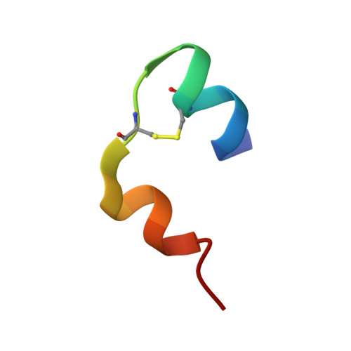
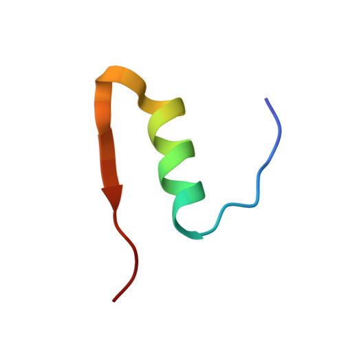
The structure of T6 human insulin at 1.0 A resolution.

Smith, G.D.Pangborn, W.A.Blessing, R.H.

(2003) Acta Crystallogr D Biol Crystallogr 59: 474-482

  • DOI: https://doi.org/10.1107/s0907444902023685
  • Primary Citation Related Structures: 
    1MSO

  • PubMed Abstract: 

    The structure of T(6) human insulin has been determined at 120 K at a resolution of 1.0 A and refined to a residual of 0.183. As a result of cryofreezing, the first four residues of the B chain in one of the two crystallographically independent AB monomers in the hexameric [Zn(1/3)(AB)(2)Zn(1/3)](3) complex undergo a conformational shift that displaces the C(alpha) atom of PheB1 by 7.86 A relative to the room-temperature structure. A least-squares superposition of all backbone atoms of the room-temperature and low-temperature structures yielded a mean displacement of 0.422 A. Omitting the first four residues of the B chain reduced the mean displacement to 0.272 A. At 120 K, nine residues were found to exhibit two discrete side-chain conformations, but only two of these residues are in common with the seven residues found to have disordered side chains in the room-temperature structure. As a result of freezing, the disorder observed at room temperature in both ArgB22 side chains is eliminated. The close contact between pairs of O( epsilon 2) atoms in GluB13 observed at room temperature is maintained at cryotemperature and suggests that a carboxylate-carboxylic acid centered hydrogen bond exists [-C(=O)-O.H.O-C(=O)-] such that the H atom is equally shared between the two partially charged O atoms.


  • Organizational Affiliation
    • Structural Biology and Biochemistry, Hospital for Sick Children, 555 University Avenue, Toronto, Ontario M5G 1X8, Canada. gdsmith@sauron.psf.sickkids.on.ca

Macromolecule Content 

  • Total Structure Weight: 11.77 kDa 
  • Atom Count: 1,103 
  • Modeled Residue Count: 102 
  • Deposited Residue Count: 102 
  • Unique protein chains: 2

Macromolecules

Find similar proteins by:  Sequence   |   3D Structure  
Entity ID: 1
MoleculeChains  Sequence LengthOrganismDetailsImage
Insulin A-Chain
A, C
21N/AMutation(s): 0 
UniProt & NIH Common Fund Data Resources
Find proteins for P01308 (Homo sapiens)
Explore P01308 
Go to UniProtKB:  P01308
PHAROS:  P01308
GTEx:  ENSG00000254647 
Entity Groups
Sequence Clusters30% Identity50% Identity70% Identity90% Identity95% Identity100% Identity
UniProt GroupP01308
Sequence Annotations
Expand
Reference Sequence
Find similar proteins by:|  3D Structure
Entity ID: 2
MoleculeChains  Sequence LengthOrganismDetailsImage
Insulin B-Chain
B, D
30N/AMutation(s): 0 
UniProt & NIH Common Fund Data Resources
Find proteins for P01308 (Homo sapiens)
Explore P01308 
Go to UniProtKB:  P01308
PHAROS:  P01308
GTEx:  ENSG00000254647 
Entity Groups
Sequence Clusters30% Identity50% Identity70% Identity90% Identity95% Identity100% Identity
UniProt GroupP01308
Sequence Annotations
Expand
Reference Sequence

Small Molecules

Experimental Data & Validation

Experimental Data

  • Method: X-RAY DIFFRACTION
  • Resolution: 1.00 Å
  • R-Value Free:  0.201 (Depositor) 
  • R-Value Work:  0.183 (Depositor) 
  • R-Value Observed: 0.183 (Depositor) 
Space Group: H 3
Unit Cell:
Length ( Å )Angle ( ˚ )
a = 81.286α = 90
b = 81.286β = 90
c = 33.714γ = 120
Software Package:
Software NamePurpose
SORTAVdata reduction
CNSrefinement
SMARTdata reduction
SORTAVdata scaling

Structure Validation

View Full Validation Report


Entry History 

Deposition Data

Revision History  (Full details and data files)

  • Version 1.0: 2003-03-04
    Type: Initial release
  • Version 1.1: 2008-04-28
    Changes: Version format compliance
  • Version 1.2: 2011-07-13
    Changes: Derived calculations, Version format compliance
  • Version 1.3: 2017-10-11
    Changes: Refinement description
  • Version 1.4: 2024-10-16
    Changes: Data collection, Database references, Derived calculations, Refinement description, Structure summary