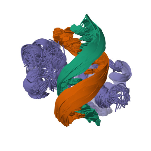
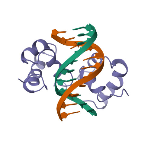
1MSF

SOLUTION STRUCTURE OF A SPECIFIC DNA COMPLEX OF THE MYB DNA-BINDING DOMAIN WITH COOPERATIVE RECOGNITION HELICES


Experimental Data Snapshot

  • Method: SOLUTION NMR
  • Conformers Submitted: 25 

wwPDB Validation   3D Report Full Report


This is version 1.4 of the entry. See complete history


Literature

Solution structure of a specific DNA complex of the Myb DNA-binding domain with cooperative recognition helices.

Ogata, K.Morikawa, S.Nakamura, H.Sekikawa, A.Inoue, T.Kanai, H.Sarai, A.Ishii, S.Nishimura, Y.

(1994) Cell 79: 639-648

  • DOI: https://doi.org/10.1016/0092-8674(94)90549-5
  • Primary Citation of Related Structures:  
    1MSE, 1MSF

  • PubMed Abstract: 

    The DNA-binding region of Myb consists of three imperfect tandem repeats (R1, R2, and R3). We have determined the solution structure of a specific DNA complex of the minimum DNA-binding domain (R2R3) by heteronuclear multidimensional NMR. Both R2 and R3 contain three helices, and the third helix in each is found to be a recognition helix. R2 and R3 are closely packed in the major groove, so that the two recognition helices contact each other directly to bind to the specific base sequence, AACNG cooperatively; this is a significant arrangement of recognition helices. The three key base pairs in this sequence are specifically recognized by Asn-183 (R3), Lys-182 (R3), and Lys-128 (R2). In contrast, R1 has no specific interactions with DNA from our NMR study of the DNA complex of the full DNA-binding domain (R1R2R3).


  • Organizational Affiliation

    Graduate School of Integrated Science, Yokohama City University, Japan.


Macromolecules

Find similar proteins by:  (by identity cutoff)  |  3D Structure
Entity ID: 3
MoleculeChains Sequence LengthOrganismDetailsImage
C-Myb DNA-Binding Domain105Mus musculusMutation(s): 0 
UniProt & NIH Common Fund Data Resources
Find proteins for P06876 (Mus musculus)
Explore P06876 
Go to UniProtKB:  P06876
IMPC:  MGI:97249
Entity Groups  
Sequence Clusters30% Identity50% Identity70% Identity90% Identity95% Identity100% Identity
UniProt GroupP06876
Sequence Annotations
Expand
  • Reference Sequence

Find similar nucleic acids by:  Sequence   |   3D Structure  

Entity ID: 1
MoleculeChains LengthOrganismImage
DNA (5'-D(*CP*CP*TP*AP*AP*CP*TP*GP*AP*CP*AP*CP*AP*CP*AP*T)-3')16N/A
Sequence Annotations
Expand
  • Reference Sequence

Find similar nucleic acids by:  Sequence   |   3D Structure  

Entity ID: 2
MoleculeChains LengthOrganismImage
DNA (5'-D(*AP*TP*GP*TP*GP*TP*GP*TP*CP*AP*GP*TP*TP*AP*GP*G)-3')16N/A
Sequence Annotations
Expand
  • Reference Sequence
Experimental Data & Validation

Experimental Data

  • Method: SOLUTION NMR
  • Conformers Submitted: 25 

Structure Validation

View Full Validation Report


Entry History 

Deposition Data

Revision History  (Full details and data files)

  • Version 1.0: 1995-03-31
    Type: Initial release
  • Version 1.1: 2008-03-24
    Changes: Version format compliance
  • Version 1.2: 2011-07-13
    Changes: Version format compliance
  • Version 1.3: 2022-02-23
    Changes: Database references, Derived calculations, Other
  • Version 1.4: 2024-05-22
    Changes: Data collection